. 2024 Feb 21;11(2):231705.
doi: 10.1098/rsos.231705. eCollection 2024 Feb.

An ascomycete H4 variant with an unknown function

Affiliations

An ascomycete H4 variant with an unknown function

Michel Flipphi et al. R Soc Open Sci. .

Abstract

Histone variants leading to altered nucleosome structure, dynamics and DNA accessibility occur frequently, albeit rarely for H4. We carried out a comprehensive in silico scrutiny of fungal genomes, which revealed the presence of a novel H4 variant (H4E) in the ascomycetes, throughout the Pezizomycotina, in basal species of the Taphrinomycotina and also in the Glomeromycota. The coding cognate genes show a specific intron/exon organization, different from H4 canonical genes. H4Es diverge from canonical H4s mainly in the N- and C-terminal extensions, showing marked differences in the distribution and number of Lys and Arg residues, which may result in novel post-translational modifications. In Aspergillus nidulans (Pezizomycotina, Eurotiomycetes) the H4E variant protein level is low in mycelia. However, the encoding gene is well expressed at 37°C under nitrogen starvation. H4E localizes to the nucleus and interacts with H3, but its absence or overexpression does not result in any detectable phenotype. Deletion of only one of the of the two canonical H4 genes results in a strikingly impaired growth phenotype, which indicates that H4E cannot replace this canonical histone. Thus, an H4 variant is present throughout a whole subphylum of the ascomycetes, but with hitherto no experimentally detectable function.

Keywords: ascomycetes; chromatin; histone H4 variant.


Conflict of interest statement

We declare we have no competing interests.

Figures
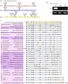
Figure 1.
Figure 1.
Overview of intron positions in H4.E genes in representative Ascomycota and RT-PCR assays to determine intron/exon organization in A. nidulans. (a) Comparison of the characteristic model intron/exon structure of the gene encoding H4E, found in Symbiotaphrina buchneri, with the intron/exon structure of the canonical H4 histones (H4.A and its paralogue H4.B). Coloured boxes refer to H4.A, H4.B and H4.E genes; vertical lines indicate intron positions in the coding sequence; texts within the triangles at the top of the vertical lines show the size in nucleotides of the cognate intron, and numbering of the intron (i.e. i1, i2, etc.). Numbers within the coloured boxes show the length of intron-separated exons. The typical gene model includes (from 5′ to 3′) a phase 2-intron, i1, separated from phase 1-intron i2 by exon 2 (29–32 nt), which in turn is separated by exon 3 (16 nt) from phase 2-intron i3, followed by an eighty 88 nt-exon 4. Phase 0-introns i4, i5, i6, and i7 are separated by exons of 60, 18, and 21 nt respectively. (b) RT-PCR assays carried out in A. nidulans to confirm the proposed intron/exon structure of hheA. mRNA samples were obtained from a wt strain grown at 25°C for 16 h on MM with 10 mM sodium nitrate (NO3) as N-source, followed by shifting the mycelia for 4 h either to MM with 5 mM ammonium tartrate as sole N-source (NH4+) or with no N-source to induce nitrogen starvation (NS), and from a PalcA:hheA:gfp strain grown on 5 mM sodium nitrate and 0.1% fructose as sole carbon source for 16 h and inducing the PalcA promoter expression by adding 50 mM ethyl methyl ketone (EMK) and further incubating for 5 h. ɣ-actin (actA) was used as control. RT-PCR products were sequenced to confirm the proposed intron/exon structure (Genbank accession MW026189). (c) Schematic presentation of conserved intron positions in genes of H4.E orthologues from representative Ascomycota. A summary of intron positions occurring in Ascomycota are shown at the top of the draw, while species-specific intron positions are listed below. Blue vertical lines denote canonical intron positions, while dotted red lines denote non-canonical intron positions. Numbers in the boxes indicate the extent of coding region between two intron positions.
Figure 2.
Figure 2.
Variation of sequence of H4Es in representatives of different fungal clades. A. oligospora (Pezizomycotina, Orbiliomycetes); N. crassa (Pezizomycotina, Sordariomycetes); S. buchneri (Pezizomycotina Xylonomycetes); T. melanosporum (Pezizomycotina, Pezizomycetes); B. cinerea and O. maius (Pezizomycotina, Leotiomycetes); A. nidulans (Pezizomycotina, Eurotiomycetes); R. irregularis, G. cerebriforme and G. margarita (Glomeromycotina, Glomeromycetes); P. fijiensis, C. zeae-maydis and Mycosphaerella sp. Ston1 (Pezizomycotina, Dothideomycetes); N. irregularis (Taphrinomycotina, Neolectomycetes); T. deformans (Taphrinomycotina, Taphrinomycetes). Red bar: core histone-fold domain; green bar: N-terminal tail basic patch; blue bar: five amino acid insertion found exclusively in the Aspergilli; orange stars: residues reported as modification targets in canonical H4s. The canonical H4 histones of Xenopus lævis, Aspergillus nidulans and Schizosaccahromyces pombe are included as references to canonical H4 conservation. Alignments carried out with MAFFT L-INS-i with default settings and visualized with Boxshade.
Figure 3.
Figure 3.
Predicted structures of selected H4Es from those aligned in figure 2. All structures were predicted with I-Tasser and drawn with VMD (see Materials and methods). The molecules were selected either for their putative independent evolutionary origin (see text) or for their striking sequence divergence. In each case the most probable model proposed by I-Tasser was chosen. (i) A. nidulans H4E (red) and Rhizophagus irregularis (Glomeromycota) H4E (blue) are superimposed to one of the Xenopus laevis canonical H4 molecules (solid green) in the nucleosome model 1kx5.pbd [32]. The inset to the right of the panel highlights a β turn present in the canonical histone but absent in both H4E models. (ii) H4E from Mycosphaerella sp. Ston1 (in orange) superimposed as below. (iii) H4E1 from Cercospora zeae-maydis (in yellow). Stars filled with cognate colours indicate the N-terminus of each protein.
Figure 4.
Figure 4.
mRNA levels of the canonical H4.1 gene (hhfA), the enigmatic variant of the H4.1 histone gene encoding the H4E variant (hheA), the H3 histone gene sharing the promoter region with H4.1 (hhtA) and the canonical H4.2 histone gene (hhfB) measured by RT-qPCR in a veA1 control strain (HZS.145). (a) Results, obtained by calculations according to the standard curve method [40], were normalized to ɣ-actin reference gene, actA. Standard deviations of three biological replicates are shown. The asterisks above the columns indicate the significance of the differences compared to the results on hhfA relative expression level. Significant differences were determined by using the Student's t-test. */**/*** indicates p < 0.05/0.01/0.001. Mycelia were grown on 10 mM sodium nitrate as the sole nitrogen source for 10 h at 37°C or 16 h at 25°C followed by transferring the mycelia to N-source-free medium (nitrogen starvation, NS), to 10 mM sodium nitrate (NO3) or to 5 mM ammonium tartrate (NH4+) and further incubated for 2 h at 37°C or 4 h for 25°C. (b) Visualization of a H4E-GFP fusion driven by its own physiological promoter at 37°C under conditions of nitrogen starvation. Black scale bars, 10 μm. BF: bright-field microscopy; DAPI: nuclei staining; GFP: GFP signal.
Figure 5.
Figure 5.
HheA::GFP colocalizes to the nucleus with a HhoA::mRFP fusion. Epifluorescence microscopy of a strain carrying a PalcA::hheA::gfp fusion when PalcA is repressed with 10 mM glucose (upper row), so no signal is seen in the GFP column, while green fluorescence is observed after PalcA induction with 10 mM threonine (second and third rows). Second column panels show the localization to the nuclei of histone H1 (HhoA) tagged with mRFP. Colocalization of GFP and RFP signals can be observed in the merge column. BF: bright-field microscopy. Growth conditions are in Materials and methods. A 100× oil immersion objective lens was used for visualization. Black scale bars, 10 µm.
Figure 6.
Figure 6.
H4E fits in a nucleosome. (a) Left: superimposition of a model of H4E obtained through I-Tasser (https://zhanggroup.org/I-TASSER/) (solid red) with a nucleosome containing a canonical H4 derived from 1KX5 [43]. The second canonical H4 molecule is shown in orange; H2A: yellow and greyish white; H2B: tan and pink; H3: dark grey and intermediate grey. Right: alignment of the amino acidic sequences of A. nidulans H4.1 and H4E. Residues that contact DNA in H4E I-Tasser model are indicated with black arrows (Arg40, Arg50, Ile51, Lys52, Asn53, Arg88, Lys89 and Leu90), while those of the canonical histone are indicated with grey arrows (Arg36, Ile 46, Lys79, Thr80). (b) Detailed view of proposed DNA-H4E contact zones. Basic residues are coloured in blue, polar residues in green, and neutral in grey. (c) Bimolecular fluorescence complementation (BiFC) microscopy to show the interaction of H4E with canonical histone H3. H3::YFPN and H4E::YFPC fusions are overexpressed from the PalcA promoter when induced with threonine (lower panels). Glucose inhibits the expression of the fusions (upper panels). BiFC YFP signals are visible in green in the nuclei, where they co-localize with mRFP-tagged histone H1 (HhoA), seen in red. BF: bright-field microscopy. Merge: co-localization of YFP and mRFP signals. Growth conditions are detailed in Materials and methods. Black scale bars, 10 µm.
Figure 7.
Figure 7.
H4E is unable to compensate for the absence of canonical H4.1. (a) Colony growth of the strains indicated in the text box below, for 48 h at 37°C on the indicated media. MVD009 is the recipient strain for the PhhfA::hheA construction, where hheA is expressed under the control of the promoter of the canonical histone H4.1 (hhfA, AN0734); yA2 hheAΔ is the recipient strain for the PalcA::hheA construction; hhfAΔ and hhfBΔ bear deletions of the canonical H4 histones H4.1 and H4.2, respectively. (b) RT-PCR to compare the expression of hheA and hhfA, in a wt strain and in a strain expressing hheA under the control of PhhfA (P::hheA). actA was amplified and used as normalization control.

Similar articles

References

    1. Luger K, Mäder AW, Richmond RK, Sargent DF, Richmond TJ. 1997. Crystal structure of the nucleosome core particle at 2.8 Å resolution. Nature 389, 251-260. (10.1038/38444) - DOI - PubMed
    1. Arents G, Burlingame RW, Wang BIC, Love WE, Moudrianakis EN. 1991. The nucleosomal core histone octamer at 3.1 Å resolution: a tripartite protein assembly and a left-handed superhelix. Proc. Natl Acad. Sci. USA 88, 10 148-10 152. (10.1073/pnas.88.22.10148) - DOI - PMC - PubMed
    1. Ehrenhofer-Murray AE. 2004. Chromatin dynamics at DNA replication, transcription and repair. Eur. J. Biochem. 271, 2335-2349. (10.1111/j.1432-1033.2004.04162.x) - DOI - PubMed
    1. Talbert PB, Henikoff S. 2017. Histone variants on the move: substrates for chromatin dynamics. Nat. Rev. Mol. Cell Biol. 18, 115-126. (10.1038/nrm.2016.148) - DOI - PubMed
    1. Clapier CR, Iwasa J, Cairns BR, Peterson CL. 2017. Mechanisms of action and regulation of ATP-dependent chromatin-remodelling complexes. Nat. Rev. Mol. Cell Biol. 18, 407-422. (10.1038/nrm.2017.26) - DOI - PMC - PubMed

LinkOut - more resources