

In vitro analysis of carotid lesions using a preliminary microwave sensor to detect vulnerable plaques: Correlation with histology, Duplex ultrasound examination, and computed tomography scanner: The Imaging and Microwave Phenotyping Assessment of Carotid stenosis Threat (IMPACT) study
- PMID: 38384784
- PMCID: PMC10879004
- DOI: 10.1016/j.jvssci.2023.100182
In vitro analysis of carotid lesions using a preliminary microwave sensor to detect vulnerable plaques: Correlation with histology, Duplex ultrasound examination, and computed tomography scanner: The Imaging and Microwave Phenotyping Assessment of Carotid stenosis Threat (IMPACT) study
Abstract
Objective: Progress in best medical treatment have made identification of best candidates for carotid surgery more difficult. New diagnostic modalities could be helpful in this perspective. Microwaves (MWs) can quantify dielectric properties (complex relative permittivity) of biological tissues and MW technology has emerged as a promising field of research for distinguishing abnormal tissues from healthy ones. We here evaluated the ability of a dedicated MW sensor developed in our laboratory to identify vulnerable carotid lesions.
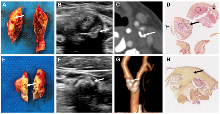
Methods: We included 50 carotid lesions in this study. The plaques were analyzed and classified preoperatively by ultrasound (US) examination, computed tomography angiography and tested postoperatively using a MW sensor. Histopathological analysis was used as a gold standard to separate vulnerable plaques (VPs) from nonvulnerable plaques (NVPs).
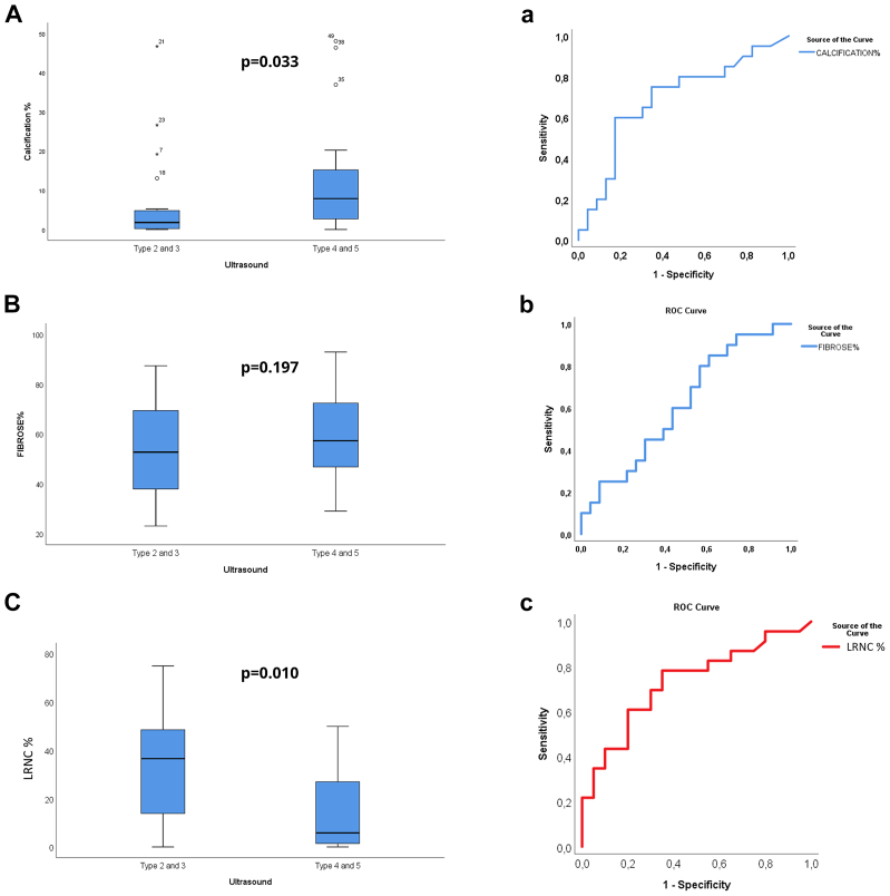
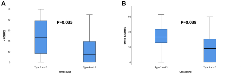
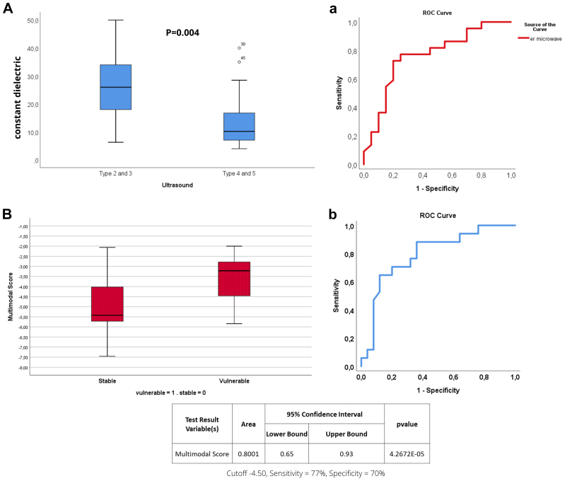
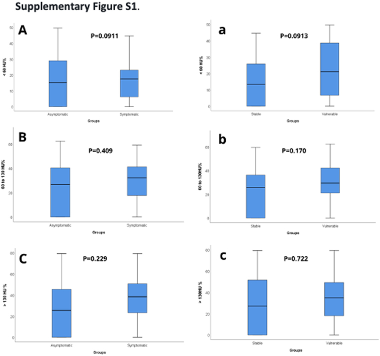
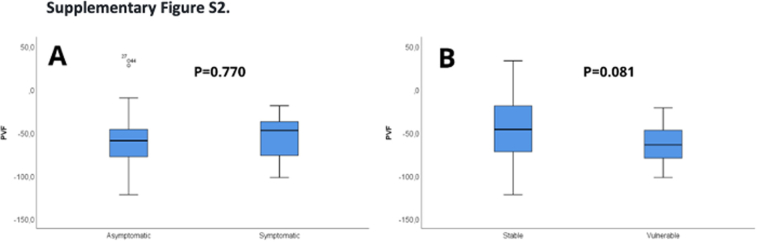
Results: VPs were more frequently types 2 or 3 plaques (on US examination), had a greater proportion of low (<60 Hounsfield unit) and moderate (60-130 Hounsfield unit) attenuation components (computed tomography angiography) and displayed higher dielectric constant values (MW) than NVPs, which had an opposite profile. NVPs were more frequently asymptomatic plaques compared with VPs (P = .035). Multivariate analysis showed that US examination and MW identified VPs with a sensitivity of 77% and a specificity of 76% (cutoff value, -0.045; area under the curve, 0.848; P < .0001).
Conclusions: We found that the presence of types 2 to 3 (on US examination) and high dielectric constant plaques in vitro was highly indicative of a VP based on histological analysis. Further studies are needed to determine the potential of MW to identify the most dangerous asymptomatic carotid lesions.
Keywords: Carotid plaque stability; Duplex US; Microwave; Risk assessment; Stroke.
© 2024 Published by Elsevier Inc. on behalf of the Society for Vascular Surgery.
Conflict of interest statement
None.
Figures
Similar articles
-
Carotid Artery Plaque Vulnerability Assessment Using Noninvasive Ultrasound Elastography: Validation With MRI.AJR Am J Roentgenol. 2017 Jul;209(1):142-151. doi: 10.2214/AJR.16.17176. AJR Am J Roentgenol. 2017. PMID: 28639927
-
Neutrophil Gelatinase Associated Lipocalin (NGAL) for Identification of Unstable Plaques in Patients with Asymptomatic Carotid Stenosis.Eur J Vasc Endovasc Surg. 2019 Jun;57(6):768-777. doi: 10.1016/j.ejvs.2018.12.029. Epub 2019 Jun 1. Eur J Vasc Endovasc Surg. 2019. PMID: 31164272
-
Identification of vulnerable carotid plaque with histologically validated CT-derived plaque maps.Br J Radiol. 2023 Jul;96(1147):20220982. doi: 10.1259/bjr.20220982. Epub 2023 May 15. Br J Radiol. 2023. PMID: 37183910 Free PMC article.
-
B-mode ultrasound and spiral CT for the assessment of carotid atherosclerosis.Neuroimaging Clin N Am. 2002 Aug;12(3):421-35. doi: 10.1016/s1052-5149(02)00015-1. Neuroimaging Clin N Am. 2002. PMID: 12486830 Review.
-
[Dual source computed tomography in analysis of significance and morphology carotid plaques].Przegl Lek. 2013;70(3):118-22. Przegl Lek. 2013. PMID: 24003664 Review. Polish.
References
-
- Reiff T., Eckstein H.H., Mansmann U., et al. Carotid endarterectomy or stenting or best medical treatment alone for moderate-to-severe asymptomatic carotid artery stenosis: 5-year results of a multicentre, randomised controlled trial. Lancet Neurol. 2022;21:877–888. - PubMed
-
- Arasu R., Arasu A., Muller J. AJGP-11-2021-Clinical-Arasu-Carotid-Artery-Stenosis-WEB. 2021;50(11):821–825. - PubMed
-
- Wardlaw J., Chappell F., Best J., Wartolowska K., Berry E. Non-invasive imaging compared with intra-arterial angiography in the diagnosis of symptomatic carotid stenosis: a meta-analysis. Lancet. 2006;367:1503–1512. - PubMed
-
- Goldstein L.B., Hasselblad V., Matchar D.B., McCrory D.C. Comparison and meta-analysis of randomized trials of endarterectomy for symptomatic carotid artery stenosis. Neurology. 1995;45:1965–1970. - PubMed
LinkOut - more resources
Full Text Sources
