. 2024 Feb 5;27(3):109107.
doi: 10.1016/j.isci.2024.109107. eCollection 2024 Mar 15.

Host ZCCHC3 blocks HIV-1 infection and production through a dual mechanism

Affiliations

Host ZCCHC3 blocks HIV-1 infection and production through a dual mechanism

Binbin Yi et al. iScience. .

Abstract

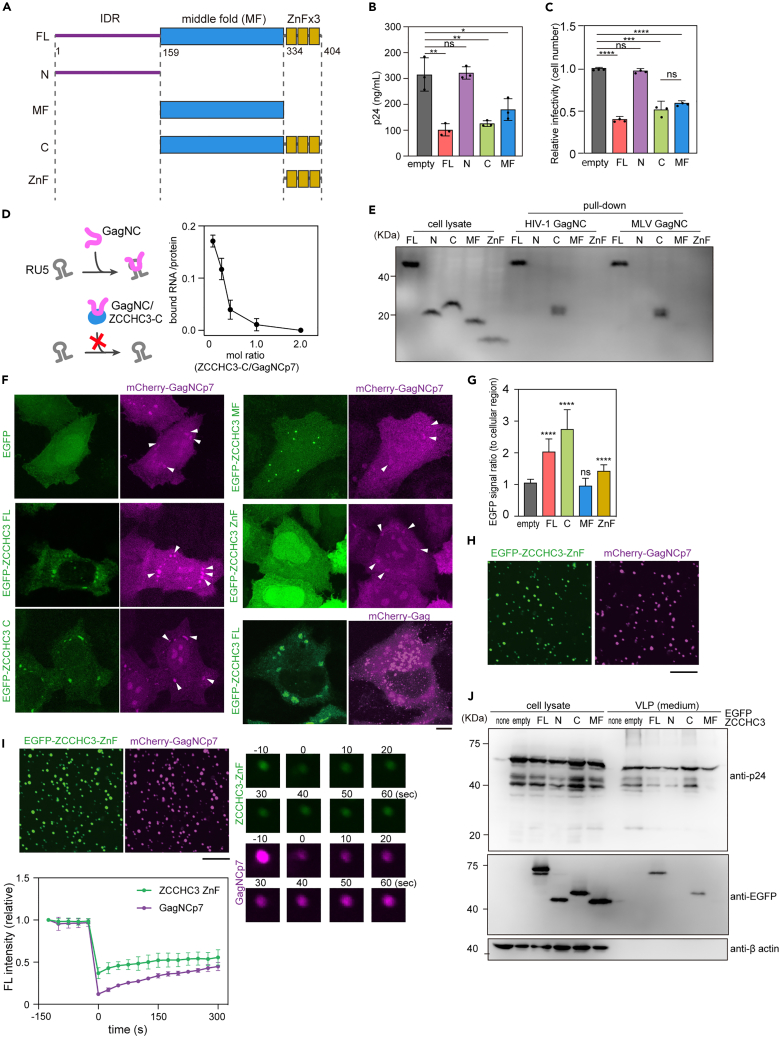
Most mammalian cells prevent viral infection and proliferation by expressing various restriction factors and sensors that activate the immune system. Several host restriction factors that inhibit human immunodeficiency virus type 1 (HIV-1) have been identified, but most of them are antagonized by viral proteins. Here, we describe CCHC-type zinc-finger-containing protein 3 (ZCCHC3) as a novel HIV-1 restriction factor that suppresses the production of HIV-1 and other retroviruses, but does not appear to be directly antagonized by viral proteins. It acts by binding to Gag nucleocapsid (GagNC) via zinc-finger motifs, which inhibits viral genome recruitment and results in genome-deficient virion production. ZCCHC3 also binds to the long terminal repeat on the viral genome via the middle-folded domain, sequestering the viral genome to P-bodies, which leads to decreased viral replication and production. This distinct, dual-acting antiviral mechanism makes upregulation of ZCCHC3 a novel potential therapeutic strategy.

Keywords: Biological sciences; Immunology; Virology.


Conflict of interest statement

J.F.H. has received research support, paid to Northwestern University, from Gilead Sciences and is a paid consultant for Merck.

Figures

None
Graphical abstract
Figure 1
Figure 1
Effect of ZCCHC3 on viral infection (A) ZCCHC3 expression in different cell types, determined in cell lysates by western blotting with an anti-ZCCHC3 antibody. HEK293T cells expressing EGFP-tagged ZCCHC3 were the positive control. The control lane contained five times less lysate than the other lanes. The panel shown here is a representative image of three independent experiments. (B) Experimental flow overview for panels in Figures 1 and 2. Producer cells (HEK293T or Lenti-X 293T) were transfected with viral plasmids and a ZCCHC3 expression plasmid, and the virions were collected from the culture medium and then analyzed as indicated. (C) Effect of ZCCHC3 on the infectivity of various viral strains. Plasmids encoding individual viral strains were introduced into Lenti-X 293T cells in the presence or absence of an HA-ZCCHC3 expression plasmid. Culture supernatant was collected 2 days after transfection and used to infect TZM-bl cells. Infectivity was determined as relative light units of luciferase 2 days after infection. The mean and standard deviation values are shown (n = 3). (D) Effect of SNPs in human ZCCHC3 gene on viral infectivity of lentiviral vectors. Lenti-X 293T cells were co-transfected with pWPI-Luc2, psPAX2-IN/HiBiT, and pMD2.G with or without an HA-ZCCHC3 expression plasmid carrying a missense mutation. Culture supernatant was collected 2 days after transfection and used to infect MT4 cells. Infectivity was determined 2 days after infection. Values relative to those for cells infected with a lentiviral vector produced with empty vector are shown as the mean ± standard deviation. (E) Effect of ZCCHC3 on viral infectivity of retroviral vectors. A plasmid encoding the indicated retrovirus and luciferase reporter gene was introduced into Lenti-X 293T cells with or without an HA-ZCCHC3 expression plasmid. Culture supernatant was collected 2 days after transfection and used to infect MT4 cells. Infectivity was determined as in (C). Values relative to those for cells harboring empty vector are shown as the mean ± standard deviation. (F) Effect of ZCCHC3 knockout on viral infection in primary CD4+ T cells. Knockout cells were generated by CRISPR-Cas9 gene editing and infected with a GFP reporter HIV-1NL4-3 virus in a spreading infection assay. Infection rates were determined by flow cytometry as percent GFP+ cells. The upper panel shows the western blot for the indicated proteins. Bar charts show cell viability (middle) and fold change in infection rates at day 5 (bottom) post-infection. A representative donor out of three shown with average of technical triplicates ±standard deviation. The results from other donors are shown in Figure S1F. (G) Effect of ZCCHC3 knockout on viral production. Lentiviruses produced in WT and ZCCHC3-knockout Lenti-X 293T cells were normalized to p24 and used to infect HeLa cells. The expression of a viral gene (EGFP) was assessed using flow cytometry (left). EGFP-positive cells were quantified (right); the mean and standard deviation values are shown (n = 3). Differences were examined using a two-tailed, unpaired Student’s t test; ∗∗∗∗p < 0.0001, ∗∗∗p < 0.001, ∗∗p < 0.01. In (C) TF2625 and TF2626, and (F), differences were examined using one-way ANOVA, followed by Tukey’s test; ∗∗∗p < 0.001, ns p ≧ 0.05.
Figure 2
Figure 2
Effect of ZCCHC3 on viral production (A) Effect of ZCCHC3 on HIV-1 viral production. A plasmid encoding HA-tagged ZCCHC3 or empty HA-vector was introduced into Lenti-X 293T cells together with an HIV-1-encoding plasmid. Virions released into the culture medium were quantified by p24 ELISA. The absolute value (top) and the value relative to that without ZCCHC3 (bottom) are shown (n = 3). (B) ZCCHC3 affects the Gag processing. Lenti-X 293T cells were co-transfected with pTF2625 plasmids in the presence or absence of HA-ZCCHC3. Equal volumes of the pelleted virions and cell lysates were analyzed via western blotting using anti-p24 and anti-HA antibodies. The total protein in the cell lysate was also analyzed via CBB staining. (C) Effect of ZCCHC3 on infectivity of ZCCHC3-loaded virions. TZM-bl cells were infected with the same amount (p24-normalized) of HIV-1NL4-3 or lentivirus with or without ZCCHC3 in the virion. Infectivity was analyzed by luciferase assay and is presented relative to that without ZCCHC3 as the mean and standard deviation (n = 5). (D) Effect of ZCCHC3 co-expression on lentivirus production. HEK293T cells were transfected with lentiviral plasmids (pLV-EGFP, psPAX2, and pMD2.G) with or without an HA-ZCCHC3 expression plasmid. Total RNA was purified from lentiviruses harvested from the culture medium and analyzed by RT-qPCR. The mean and standard deviation values are shown (n = 3). In (A), (C), and (D), differences were examined by a two-tailed, unpaired Student’s t test; ∗∗∗∗p < 0.0001, ∗∗∗p < 0.001, ∗∗p < 0.001, ∗p < 0.05.
Figure 3
Figure 3
ZCCHC3 binding to GagNC (A) ZCCHC3 binding to NCp7 of HIV-1 Gag. GST-tagged HIV-1 Gag MAp17, CAp24, NCp7 or p6 protein, or MLV Gag NC was mixed with HEK293T cell lysate containing HA-ZCCHC3 and GSH beads after treatment with RNase A. The eluted fraction was analyzed by western blotting using an anti-HA antibody. A representative image from three independent experiments is shown here. See also Figure S3A. (B) ZCCHC3 incorporated into the HIV-1 TF2625 virion. Lenti-X 293T cells were transfected with pTF2625 plasmid in the presence or absence of an EGFP-ZCCHC3 expression plasmid. Pelleted virions were analyzed via western blotting with anti-ZCCHC3, anti-p24, and anti-β-actin antibodies. (C) Lentiviral plasmids (pLV-EGFP, psPAX2, and pIIIenv3-1) (left) or a plasmid encoding HIV-1 Gag (right) were introduced into 293T cells with or without an HA-ZCCHC3 expression plasmid. Virions were harvested by centrifugation, and analyzed by immunoblotting with anti-EGFP, anti-p24, and anti-β-actin antibodies. (D) The zinc-finger motifs of GagNCp7 are not necessary for interaction with ZCCHC3. GST-tagged GagNCp7 (WT or mutant) was mixed with the cell lysate containing HA-ZCCHC3 and GSH beads. The position of the mutations (C to S) in GagNCp7 is depicted in the top panel. The bound fraction was analyzed via western blotting using an anti-HA antibody. A representative image from three independent experiments is shown here. See also Figure S3B. (E) Presence of ZCCHC3 in HIV-1 Gag VLP. COS7 cells expressing HA-ZCCHC3 and mCherry-HIV-1 Gag were fixed, stained with an anti-HA antibody, and observed using confocal laser scanning microscopy (CLSM) (left). COS7 cells expressing EGFP-ZCCHC3 and mCherry-HIV-1 Gag were fixed and observed using CLSM (right). Enlarged images are also shown at the bottom. Scale bars, 5 μm (top), 25 μm (bottom).
Figure 4
Figure 4
ZCCHC3 binding to GagNC via C-terminal domain (A) The domain structure of human ZCCHC3. IDR, MF, and zinc-finger (ZnF) domains are indicated. (B and C) Effect of C-terminal fragment of ZCCHC3 on viral production and infectivity. HEK293T cells were transfected with lentiviral vectors, and a ZCCHC3 FL, N, C, or MF expression vector or an empty vector. The resultant lentiviruses were harvested 2 days after transfection and quantified by p24 ELISA (B). HeLa cells were infected with a p24-normalized amount of harvested lentiviruses, and infectivity was quantified based on the expression of a viral gene (EGFP) using flow cytometry (C). The mean and standard deviation values from three independent experiments are shown. (D) Hypothetical mechanism of ZCCHC3 inhibition of the interaction between Gag NC and viral RNA and the effect of ZCCHC3 on the interaction between HIV-1 GagNCp7 and LTR RNA. Purified ZCCHC3 was tested in RNA pull-down assay of GagNCp7 and HIV-1 LTR, at the molar ratios indicated. A fluorescent probe was used to quantify RNA (ng) in the bound fraction, which is presented as a ratio to the amount of the bait protein (ng) in the bound fraction quantified by Coomassie brilliant blue (CBB) staining. The mean and standard deviation values from three independent experiments are shown. (E) Binding of different ZCCHC3 domains to GagNCp7. GST-tagged HIV-1 or MLV Gag NC was mixed with HEK293T cell lysate containing HA-tagged ZCCHC3 FL, N, C, MF, or ZnF fragments after treating with RNase A, and the eluted fraction was analyzed using western blotting with anti-HA antibody. See also Figure S3F. (F and G) Localization of HIV-1 GagNCp7 and ZCCHC3 in HeLa cells. HeLa cells expressing mCherry-Gag NCp7 and EGFP-tagged ZCCHC3 FL, C, MF, or ZnF fragments were fixed and observed using CLSM (F). The arrowheads indicate the cytoplasmic GagNCp7 condensate. Scale bar, 5 μm. The fluorescence intensity ratio of GagNCp7 foci and cytoplasm was quantified (n = 15) (G). (H and I) ZCCHC3 ZnF domain co-localizes with GagNCp7 in vitro. In vitro droplet assay using recombinant Hisx6-EGFP-ZCCHC3 ZnF and Hisx6-mCherry-GagNCp7. Both proteins formed droplets in the presence of 15% polyethyleneglycol (H). Scale bar, 100 μm. Both proteins co-localized in the same droplet when they were mixed together (I, top left). FRAP analysis was performed against the droplet to measure the mobility of the proteins. Representative images are presented (right). The average fluorescence intensity of the bleached region was quantified and plotted against time as a value relative to that of the pre-bleached signal. Data are presented as mean ± standard deviation from three independent measurements. Scale bar, 100 μm. (J) Incorporation of ZCCHC3 domains into Gag VLP. Plasmid encoding EGFP-tagged ZCCHC3 FL, N, C, or MF was introduced into HEK293T cells together with a plasmid encoding HIV-1 Gag. VLPs released into the culture medium were harvested and analyzed by immunoblotting with anti-GFP, anti-p24, and anti-β-actin antibodies. In (B), (C), and (G) differences were examined by a two-tailed, unpaired Student’s t test; ∗∗∗∗p < 0.0001, ∗∗∗p < 0.001, ∗∗p < 0.01, ns, p ≥ 0.05.
Figure 5
Figure 5
ZCCHC3 binding to retroviral RNA (A) 3D structure of ZCCHC3 MF region predicted by using AlphaFold2. Basic residues in the central cleft are labeled in blue. (B) ZCCHC3 MF domain binding to LTR RNAs of HIV-1 and MLV. GST-tagged ZCCHC3 MF or GST was mixed with ssRNA, dsDNA, or ssDNA of HIV-1 LTR (R-U5), MLV LTR, or a coding region of Gag. Nucleic acids in the bound fraction were quantified using a fluorescent probe, and the amount is presented as a molecular ratio to the bait protein. The mean and standard deviation values from three independent experiments are shown. (C) EMSA analysis of ZCCHC3–HIV-1 genome interaction. Different amounts of purified ZCCHC3 were incubated with ssRNA, dsDNA, and ssDNA (0.1 pmol, prepared as in B) and analyzed. (D) Binding of ZCCHC3 domains to LTR, stem-loops of the LTR and RRE. RNA pull-down assay was performed with HIV-1 LTR (R-U5), SL1, SL2, SL3 RNA or HIV-1 RRE and GST-tagged ZCCHC3 FL or MF as described in (B). The amount of bound RNA is presented as the molecular ratio to the bait protein, with the mean and standard deviation values from three independent experiments shown. (E) Schematic illustration of HIV-1 LTR (R-U5) secondary structure. (F) Binding of ZCCHC3 MF domain WT and basic amino acid mutants to the stem-loops of HIV-1 LTR (R-U5). RNA pull-down assay was performed with the HIV-1 LTR (R-U5), and GST-ZCCHC3 MF (WT or mutants), as described in (D). The amount of bound RNA is presented as the molecular ratio to the bait protein, with the mean and standard deviation values from three independent experiments shown. (G) ZCCHC3 suppresses the expression of LTR-containing genes. The promotor constructs are depicted (left). HIV-1 LTR (R-U5) was inserted upstream or downstream of the EGFP ORF. A fragment of the HIV-1 Gag gene (181 bp) was used as a control. Some constructs carry HIV-1 LTR (full length) or chicken β-actin promoter in place of the CMV promoter. The EGFP reporter constructs were introduced into HEK293T cells with or without an HA-ZCCHC3 expression plasmid, and EGFP fluorescence was quantified by flow cytometry. Quantification of EGFP-expressing cells as the ratio of signal between cells with (+) and without (−) HA- ZCCHC3 is shown (right). The mean and standard deviation values from three independent experiments are shown. Differences in B, D, F and G were examined by a two-tailed, unpaired Student’s t test. ∗∗∗p < 0.001, ∗∗p < 0.01, ∗p < 0.05; ns, p ≥ 0.05.
Figure 6
Figure 6
ZCCHC3 sequestration of viral RNA in P-body (A–C) ZCCHC3 localization in the P-body. HEK293T cells were non-transfected or transfected with EGFP-ZCCHC3-encoding plasmid, lentiviral plasmid, or a plasmid carrying HIV-1 LTR, and immuno-stained with anti-LSM14A and anti-ZCCHC3 antibodies. Representative images are shown (A). Scale bar, 5 μm. The number of LSM14A-positive foci per cell was counted (20 cells in each condition) and is summarized in (B). The fraction of ZCCHC3-containing P-bodies was counted and is summarized in (C). (D and E) BioID analysis of 293T cells stably expressing TurboID-fused ZCCHC3. P-body proteins were extracted from the set of identified proteins using GO terms and are shown for three independent experiments (D). The original data of mass spectrometry is provided in Table S2. The total numbers of proteins identified in individual experiments are summarized in a Venn diagram (E). Fold enrichment of P-body proteins is shown as the mean and standard deviation (n = 3; E, right bottom panel). (F) The role of ZCCHC3 MF domain in P-body localization. HEK293T cells expressing EGFP-tagged fragments (N, MF, C) of ZCCHC3 or ZCCHC3 FL carrying R168/248S mutations were immuno-stained with an anti-LSM14A antibody and observed using CLSM. Scale bars, 5 μm (left panels), 1 μm (right panels). (G) Proposed mechanism of HIV-1 suppression by ZCCHC3. The 5′ LTR region of nuclear-exported HIV-1 genomic RNA is recognized by the ZCCHC3 MF, and sequestered in the P-bodies, which impairs virion maturation. ZCCHC3 also binds to HIV-1 Gag NCp7, which leads to ZCCHC3 incorporation into the virion and promotes the antiviral function of ZCCHC3 during subsequent infection. Differences in (B), (C), and (E) were examined by a two-tailed, unpaired Student’s t test. ∗∗∗∗p < 0.0001, ∗∗∗p < 0.001, ∗p < 0.05.

References

    1. Fauci A.S. Host factors and the pathogenesis of HIV-induced disease. Nature. 1996;384:529–534. doi: 10.1038/384529a0. - DOI - PubMed
    1. Ishii K.J., Coban C., Kato H., Takahashi K., Torii Y., Takeshita F., Ludwig H., Sutter G., Suzuki K., Hemmi H., et al. A Toll-like receptor–independent antiviral response induced by double-stranded B-form DNA. Nat. Immunol. 2006;7:40–48. doi: 10.1038/ni1282. - DOI - PubMed
    1. Ghimire D., Rai M., Gaur R. Novel host restriction factors implicated in HIV-1 replication. J. Gen. Virol. 2018;99:435–446. doi: 10.1099/jgv.0.001026. - DOI - PubMed
    1. Smith H.C., Bennett R.P., Kizilyer A., McDougall W.M., Prohaska K.M. Functions and regulation of the APOBEC family of proteins. Semin. Cell Dev. Biol. 2012;23:258–268. doi: 10.1016/j.semcdb.2011.10.004. - DOI - PMC - PubMed
    1. Albin J.S., Harris R.S. Interactions of host APOBEC3 restriction factors with HIV-1 in vivo: Implications for therapeutics. Expet Rev. Mol. Med. 2010;12:e4. doi: 10.1017/S1462399409001343. - DOI - PMC - PubMed