. 2023 Nov 27;27(1):2747.
doi: 10.4102/sajr.v27i1.2747. eCollection 2023.

Imaging spectrum of spinal dysraphism: A diagnostic challenge

Affiliations

Imaging spectrum of spinal dysraphism: A diagnostic challenge

Mohit K Shrivastva et al. SA J Radiol. .

Abstract

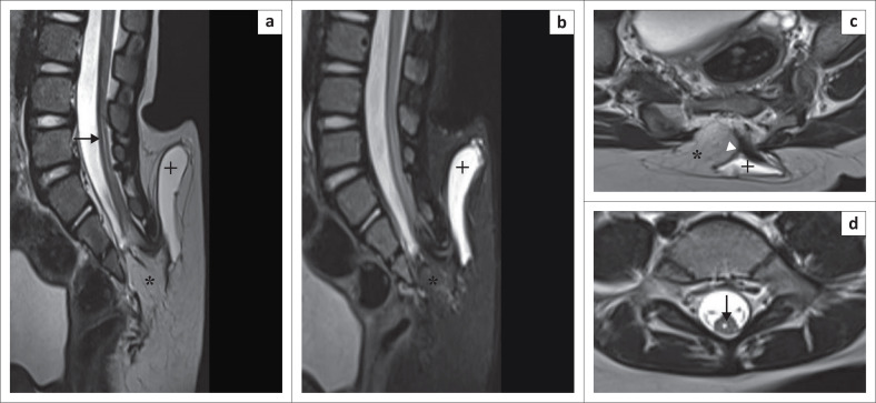
Spinal dysraphism (SD) is a collective term for congenital malformations of the spine and spinal cord. It includes a wide range of congenital anomalies resulting from aberrations in the stages of gastrulation, primary neurulation and secondary neurulation. Spinal dysraphism may lead to neurological impairment of varying severity including weakness of the extremities, incontinence of bowel and bladder, sexual dysfunction, among others. Diagnosis of SDs is quite challenging because of its wide spectrum and complex cascade of embryologic events. Knowledge of normal embryology and proper understanding of imaging features of SD are important for early accurate diagnosis.

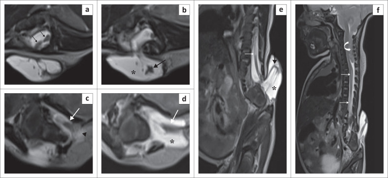
Contribution: This series of five cases describes the imaging spectrum of spinal dysraphism and highlights the embryological basis for their development, which could facilitate early correct diagnosis, surgical planning and reduced morbidity related to these malformations. It also includes an extremely rare case of complex spinal dysraphism (Type II diastematomyelia with right hemimyelomeningocoele and left hemilipomyelomeningocoele) with Chiari II malformation.

Keywords: caudal regression syndrome; diastematomyelia; dorsal dermal sinus; hemimyelomeningocoele; lipomyelocoele; lipomyelomeningocoele; myelomeningocoele; spinal dysraphism.


Conflict of interest statement

The authors declare that they have no personal or financial relationship that may have inappropriately influenced the writing of this article.

Figures

FIGURE 1
FIGURE 1
Lipomyelomeningocoele. Sagittal T2-weighted image (T2WI) (a) and sagittal short tau inversion recovery (STIR) (b) demonstrate a low-lying spinal cord tethered to a lipoma (*). Axial T2WI (c) shows protrusion of neural tissue (white arrowhead) and an enlarged cerebrospinal fluid (CSF) space (+) through the bony defect in the posterior elements. The cord–lipoma interface is present outside the spinal canal. The overlying skin is intact. Syrinx formation (black solid arrow) is noted (a, d) at the L5 and S1 vertebral levels.
FIGURE 2
FIGURE 2
Lipomyelocoele. Sagittal T2-weighted image (T2WI) (a) shows a low-lying spinal cord tethered to a lipoma (*). The lipoma is extending into the spinal canal and is continuous with the subcutaneous fat posteriorly on sagittal T2WI (b), sagittal short tau inversion recovery (STIR) (c) and axial T2WI (d). Sagittal and axial T2WI (b, d) show the cord–lipoma interface (black solid arrow) at the level of the neural arches, without widening of the subarachnoid space. Volume rendered CT image (e) demonstrates a deformed sacrum.
FIGURE 3
FIGURE 3
Type II diastematomyelia with a right hemimyelomeningocoele, left hemilipomyelomeningocoele and Arnold Chiari II malformation. Axial T2-weighted image (T2WI) (a) shows the splitting of spinal cord (diastematomyelia) (thin black arrows). Axial and sagittal T2WI (b & e) show herniation of the right hemicord (solid black arrows) along with an enlarged subarachnoid space (*) through the right posterior bony defect with no overlying skin or subcutaneous tissue (right hemimyelomeningocoele). Axial T2WI (c) shows herniation of the left hemicord (solid white arrow) through the left posterior bony defect with tethering to a subcutaneous lipomatous mass (black arrowhead). Axial T2WI (d) shows protrusion of the neural tissue and an enlarged CSF space (*) through the bony defect (left hemilipomyelomeningocoele). Sagittal T2WI (f) shows herniation of the cerebellum (white curved arrow) into the spinal canal through the foramen magnum and syrinx formation (thin white arrow).
FIGURE 4
FIGURE 4
Type I caudal regression syndrome. Sagittal T2-weighted image (T2WI) (a) and radiographs (d, e) show non-visualisation of the sacrum (beyond S1) and coccyx with a L1–L2 block vertebra (+). There is mild anterior wedging of the L3 vertebra. Coronal short tau inversion recovery (STIR) (c) shows a vertical midline cleft (thin black arrow) in the S1 vertebral body. Sagittal T2WI (a) shows attenuation of the spinal cord (solid white arrow) from the T10 to T12 vertebral levels and a thickened conus medullaris (*) with abrupt termination at the lower end of the block L1–L2 vertebra. The nerve roots follow an atypical course having a ‘double-bundle shape’ (a, b) (thin white arrows).
FIGURE 5
FIGURE 5
Dorsal dermal sinus. Sagittal T2-weighted image (T2WI) (a), T1WI (b), short tau inversion recovery (STIR) (c) and axial T2WI (d) show a sinus track (thin white arrow) extending from the skin surface to the thoracic spinal cord through a posterior bony defect (thin black arrows in d) at the T3 vertebral level. A heterogeneous lobulated intradural extramedullary collection (*) is causing significant spinal cord compression with associated spinal cord oedema/myelomalacia (solid white arrow).
FIGURE 6
FIGURE 6
Gastrulation (a–c): (a) The hypoblast (orange cells) and the epiblast (blue cells) form the bilaminar embryonic disc. (b) Hypoblast forms the endoderm after being displaced caudally by cells (green cells) arising from the primitive pit (red circle) entering between the epiblast and hypoblast. (c) Mesoderm (magenta) and notochord (magenta circle) are formed by cells migrating bilaterally above the endoderm. Trilaminar disc and notochord are formed. Primary neurulation (d–f): (d) Formation of the neural plate is stimulated by the notochord in the dorsal midline of the ectoderm. (e) Two neural folds with neural groove in between are formed. (f) The neural folds fuse in the midline and detach from the cutaneous ectoderm, forming the neural tube. Secondary neurulation (g–i): (g) Caudal to the primary neural tube, tail bud (red) is formed by a solid mass of totipotent cells. (h) Secondary neural tube is formed by internal cavitation in the tail bud. (i) Secondary neural tube joins the primary neural tube (deep blue) creating a continuous structure.
FIGURE 7
FIGURE 7
Clinical-radiologic classification of spinal dysraphisms.

References

    1. Trapp B, 4Freddi TdAL, Hans MdOM, et al. . A practical approach to diagnosis of spinal dysraphism. Radiographics. 2021;41(2):559–575. 10.1148/rg.2021200103 - DOI - PubMed
    1. Chellathurai A, Rajendran K. Spinal dysraphisms: A new anatomical – Clinicoradiological classification. Indian J Radiol Imaging. 2022;31(4):809–829. 10.1055/s-0041-1741100 - DOI - PMC - PubMed
    1. Singh N, Singh DK, Kumar R. Diastematomyelia with hemimyelomeningocele: An exceptional and complex spinal dysraphism. J Pediatr Neurosci. 2015;10(3):237–239. 10.4103/1817-1745.165665 - DOI - PMC - PubMed
    1. Jans L, Vlummens P, Van Damme S, Verstraete K, Abernethy L. Hemimyelomeningocele: A rare and complex spinal dysraphism. JBR-BTR. 2008;91(5):198–199. - PubMed
    1. Kumar J, Afsal M, Garg A. Imaging spectrum of spinal dysraphism on magnetic resonance: A pictorial review. World J Radiol. 2017;9(4):178–190. 10.4329/wjr.v9.i4.178 - DOI - PMC - PubMed

LinkOut - more resources