Review
. 2023 Nov 28;27(1):2707.
doi: 10.4102/sajr.v27i1.2707. eCollection 2023.

Superior labrum anterior to posterior lesions: Part 2 - Classification with arthroscopic correlation

Affiliations
Review

Superior labrum anterior to posterior lesions: Part 2 - Classification with arthroscopic correlation

Peter Mercouris et al. SA J Radiol. .

Abstract

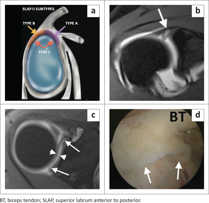
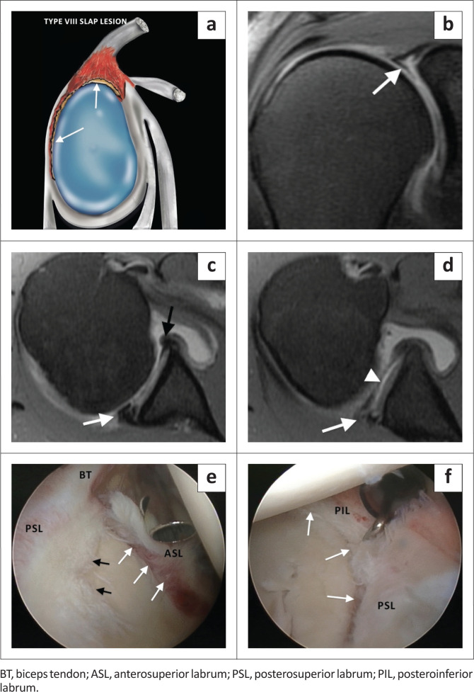
The glenoid labrum deepens the glenoid fossa and allows for the attachment of the long head of the biceps tendon and glenohumeral ligaments, contributing to the stability of the glenohumeral joint. The superior labrum is a common site of labral injury. The acronym SLAP (superior labrum anterior to posterior or anteroposterior) lesion was introduced by Snyder and colleagues in 1990 to describe superior labral tears based on arthroscopic evaluation. This original classification has since been expanded, and there are currently 10 types of SLAP lesions. The article will describe and illustrate the 10 types of SLAP lesions by means of colour illustrations, MRI images and correlative arthroscopy images. A practical approach to the assessment of SLAP lesions will be recommended.

Contribution: The illustrated review functions as a crucial radiological guide for both radiologists and orthopaedic surgeons. The combination of illustrations, MR and correlative arthroscopic images enhances the comprehensive understanding of labral pathology. The value of the review lies in the presentation of imaging findings and classification, coupled with findings on arthroscopy. This understanding is vital in guiding orthopaedic management for patients, ensuring appropriate treatment strategies.

Keywords: MRI; SLAP lesion or tear; anatomic variants; arthroscopy; glenoid labrum; shoulder.


Conflict of interest statement

The authors declare that they have no financial or personal relationships that may have inappropriately influenced them in writing this article.

Figures

FIGURE 1
FIGURE 1
A SLAP type I lesion. (a) Colour illustration. (b) Degenerative fraying (white arrow) of the superior labrum. Note the high attachment of the inferior glenohumeral ligament (black arrow). (c) FS T1-weighted MR arthrogram coronal image demonstrating an articular cartilage defect (between the black arrows) in the superomedial portion of the humeral head. (d) Arthroscopic confirmation of fraying of the superior labrum (white arrow). (e) Arthroscopic confirmation of a chondral fracture (white arrows).
FIGURE 2
FIGURE 2
Sublabral recess versus SLAP tear. (a) Colour illustration of a sublabral recess or sulcus – white line (or recess) is medially oriented. (b) FS T1 coronal oblique arthrogram demonstrating medial orientation of contrast-filled recess. (c) Colour illustration of SLAP tear – white line of tear is laterally oriented. (d) FS T1 coronal oblique arthrogram demonstrating the lateral orientation of the contrast-filled tear.
FIGURE 3
FIGURE 3
‘Oreo cookie’ sign. (a) Illustration showing a single ‘Oreo cookie’ in a sublabral recess (black arrow). (b) Illustration showing a double ‘Oreo cookie’ sign in a SLAP III tear. Long black arrow – sublabral recess, short black arrow – contrast (or fluid) filled defect of the displaced labral tear. (c) FS T1-weighted MR arthrogram coronal oblique image reflecting the illustration in (b) in this patient with an arthroscopically proven type III SLAP tear. The long black arrow represents the sublabral recess and the short black arrow the contrast-filled defect of the displaced labral tear (black arrowhead).
FIGURE 4
FIGURE 4
A SLAP type II lesion. (a) Colour illustration. (b) FS T1-weighted (T1W) MR arthrogram coronal oblique image demonstrating an enlarged (> 5 mm) globular appearing sublabral contrast collection indicative of a superior labral tear (black arrow). (c) FS T1W MR arthrogram coronal oblique image depicting a laterally oriented contrast collection (black arrow), a feature of a labral tear. (d) FS T1W MR arthrogram coronal oblique image posterior to image (c) confirms the extension of the superior labral tear posterior to the biceps anchor. White arrow shows incidental calcification in the infraspinatus tendon. (e) Arthroscopic (posterior portal) confirmation of a type II lesion. The probe elevates the torn and detached superior labrum (white arrows).
FIGURE 5
FIGURE 5
The SLAP II lesion subtypes. (a) Colour illustration demonstrating the SLAP II subtypes: (1) Type A – anterior SLAP II lesion (purple shading and arrow). (2) Type B – posterior SLAP II lesion (orange shading and arrow). (3) Type C – classic type II SLAP lesion with anterior and posterior extension. (b) FS T1-weighted (T1W) MR arthrogram coronal oblique image demonstrating a superior labral tear with biceps anchor involvement (white arrow). (c) FS T1W MR arthrogram axial image depicting the full anterior to posterior extent (white arrows) of the superior labral tear and detachment (white arrowheads) indicating a type C SLAP II tear. (d) Arthroscopic (posterior portal) confirmation of the SLAP II tear (white arrows).
FIGURE 6
FIGURE 6
A SLAP type III lesion. (a) Lateral colour illustration of a SLAP III lesion. (b) FS T1-weighted (T1W) MR arthrogram coronal oblique image demonstrating an inferiorly displaced superior labrum (white arrow) consistent with a bucket handle tear but with an intact biceps anchor (black arrow). (c) Arthroscopic probe displacing the bucket handle component inferiorly (black arrows).
FIGURE 7
FIGURE 7
A SLAP type IV lesion. (a) Lateral colour illustration of a SLAP IV tear. (b) FS T1-weighted (T1W) MR arthrogram coronal image showing inferior displacement of the torn superior labrum (black arrow) and involvement of the biceps anchor (white arrow). (c) Arthroscopic image confirming the bucket handle tear extending into the biceps anchor (black arrows).
FIGURE 8
FIGURE 8
‘Triple structure’ sign. (a) FS T1-weighted (T1W) MR arthrogram coronal image showing inferior displacement of the torn superior labrum (white arrow) and involvement of the biceps anchor (black arrow) of an arthroscopically confirmed SLAP type IV tear. (b) FS T1W MR arthrogram sagittal oblique image demonstrating the ‘three structure’ sign. Black arrow depicts the biceps tendon and white arrows depict the bucket handle fragments.
FIGURE 9
FIGURE 9
A SLAP type V lesion. (a) Lateral colour illustration of a SLAP V tear. (b) FS T2-weighted (T2W) coronal oblique (post arthrogram) image demonstrating abnormal superior labral morphology (white arrow) and anterosuperior labral detachment (black arrow). (c) FS T1-weighted (T1W) post arthrogram axial image showing the anteroinferior labral tear (white arrow) of a Bankart lesion. (d) FS T1W post arthrogram sagittal image depicting detachment of entire anterior labrum (white arrows). (e) Arthroscopic (posterior portal) image confirming the superior labral tear (black arrow) and detachment of anterosuperior labrum (white arrows). (f) Arthroscopic image showing the tear extending to involve the entire anterior labrum (white arrows).
FIGURE 10
FIGURE 10
A SLAP type VI lesion. (a) Lateral colour illustration of a SLAP VI tear. (b) FS T1-weighted (T1W) MR arthrogram coronal image displaying the displaced labrum (white arrow). (c) FS T1 MR arthrogram sagittal image displaying the anteriorly displaced flap of labral tissue (black arrows). (d) Arthroscopic (posterior portal) image confirming the anterior flap (white arrows).
FIGURE 11
FIGURE 11
A SLAP type VII lesion. (a) Lateral colour illustration of a SLAP VII tear. (b) FS T1-weighted (T1W) MR arthrogram axial image showing a superior labral tear extending from anterior to posterior (white arrows). (c) FS T1W MR arthrogram sagittal image demonstrating extension of this superior labral tear (black arrow) into the middle glenohumeral ligament (white arrows) in this arthroscopically confirmed SLAP VII tear.
FIGURE 12
FIGURE 12
A SLAP type VIII lesion. (a) Lateral colour illustration of a SLAP VIII tear. (b) FS T1-weighted (T1W) MR arthrogram coronal oblique image showing the complex tear pattern of the superior labrum including the biceps anchor (white arrow). (c) FS T1W MR arthrogram axial image below the level of the coracoid process depicting the posteroinferior extension of the labral tear. White arrow – torn posterior labrum and black arrow – normal anterior labrum. (d) FS T1W MR arthrogram axial image at the level of the inferior joint indicating that the tear extends to involve the inferior labrum at the 6 o’clock position. White arrow denotes the torn posteroinferior labrum and white arrowhead the detachment of the inferior labrum. (e) Arthroscopy confirming the superior labral tear with anterior (white arrows) and posterior (black arrows) extension. (f) Arthroscopic view with white arrows depicting the torn and detached posterosuperior (PSL) and posteroinferior labrum (PIL).
FIGURE 13
FIGURE 13
A SLAP type VIII lesion. (a) FS T1-weighted (T1W) MR arthrogram coronal oblique image demonstrating a superior labral tear (white arrow). (b) FS T1W MR arthrogram axial image depicting a tear of the posterior labrum with gadolinium-based contrast filling of the paralabral cysts (white arrows). (c) FS T1W MR arthrogram axial image at the level of the inferior margin of the subscapularis tendon showing the inferior extent of the tear of the posterior labrum (white arrow). (d) FS T1W MR arthrogram coronal oblique image demonstrating a paralabral cyst filling with only a very small amount of gadolinium-based contrast (white arrow). (e) T2-weighted (T2W) coronal oblique image depicting the true size of the paralabral cyst, highlighting the importance of some form of T2W sequence as part of the imaging protocol. (f) Arthroscopic confirmation of a SLAP VIII tear. White arrows indicate the superior labral tear. Black arrow indicates the arthroscopic probe overlying the biceps tendon. (g) Arthroscopic image demonstrating the posterior extension of the tear (white arrows) to involve the posterosuperior (PSL) and posteroinferior labrum (PIL).
FIGURE 14
FIGURE 14
A SLAP type IX lesion. (a) Lateral colour illustration of a SLAP IX lesion. (b–e) Axial images (FS T1-weighted (T1W) MR arthrogram) from superior to inferior through the entire labrum displaying the detachment of both the anterior and posterior labrum (white arrows). (f–h) Arthroscopic confirmation of a circumferential labral tear (white arrows) in this 29-year-old professional rugby player.
FIGURE 15
FIGURE 15
A SLAP type X lesion. (a) Lateral colour illustration of a SLAP X tear. (b) FS T1-weighted (T1W) MR arthrogram axial image depicting full anterior to posterior involvement (white arrows) of the superior labral tear with extension into the superior glenohumeral ligament (SGHL) (black arrows). (c) Arthroscopy confirming a tear of the SGHL (arrow).
FIGURE 16
FIGURE 16
Practical approach to SLAP tear diagnosis on magnetic resonance imaging.
FIGURE 17
FIGURE 17
A suggested algorithm for operative management of SLAP lesions.

References

    1. Andrews JR, Carson WG Jr, McLeod WD. Glenoid labrum tears related to the long head of the biceps. Am J Sports Med. 1985;13(5):337–341. 10.1177/036354658501300508 - DOI - PubMed
    1. Snyder SJ, Karzel RP, Del Pizzo W, et al. . SLAP lesions of the shoulder. Arthroscopy. 1990;6(4):274–279. 10.1016/0749-8063(90)90056-J - DOI - PubMed
    1. Mohana-Borges AV, Chung CB, Resnick D. Superior labral anteroposterior tear: Classification and diagnosis on MRI and MR arthrography. AJR Am J Roentgenol. 2003;181(6):1449–1462. 10.2214/ajr.181.6.1811449 - DOI - PubMed
    1. Stoller DW. The shoulder. Chapter 8. In: Stoller DW, editor. Magnetic resonance imaging in orthopaedics and sports medicine. 3rd ed. Philadelphia: Lippincott Williams & Wilkins, 2007; p. 1131–1461.
    1. Chung CB, Steinbach LS. Long bicipital tendon including superior labral anterior-posterior lesions, Chapter 6. In: Steinback LS, Tirman PFJ, Peterfy CG, Feller JF, editors. MRI of the upper extremity: Shoulder, elbow, wrist and hand. Chapter 6. Philadelphia, PA: Wolters Kluver Helath/Lippincott Williams & Wilkins, 2010; p. 337.

LinkOut - more resources