. 2024 Apr;44(4):930-945.
doi: 10.1161/ATVBAHA.123.320524. Epub 2024 Feb 22.

High-Dimensional Single-Cell Multimodal Landscape of Human Carotid Atherosclerosis

Affiliations

High-Dimensional Single-Cell Multimodal Landscape of Human Carotid Atherosclerosis

Alexander C Bashore et al. Arterioscler Thromb Vasc Biol. 2024 Apr.

Abstract

Background: Atherosclerotic plaques are complex tissues composed of a heterogeneous mixture of cells. However, our understanding of the comprehensive transcriptional and phenotypic landscape of the cells within these lesions is limited.

Methods: To characterize the landscape of human carotid atherosclerosis in greater detail, we combined cellular indexing of transcriptomes and epitopes by sequencing and single-cell RNA sequencing to classify all cell types within lesions (n=21; 13 symptomatic) to achieve a comprehensive multimodal understanding of the cellular identities of atherosclerosis and their association with clinical pathophysiology.

Results: We identified 25 cell populations, each with a unique multiomic signature, including macrophages, T cells, NK (natural killer) cells, mast cells, B cells, plasma cells, neutrophils, dendritic cells, endothelial cells, fibroblasts, and smooth muscle cells (SMCs). Among the macrophages, we identified 2 proinflammatory subsets enriched in IL-1B (interleukin-1B) or C1Q expression, 2 TREM2-positive foam cells (1 expressing inflammatory genes), and subpopulations with a proliferative gene signature and SMC-specific gene signature with fibrotic pathways upregulated. Further characterization revealed various subsets of SMCs and fibroblasts, including SMC-derived foam cells. These foamy SMCs were localized in the deep intima of coronary atherosclerotic lesions. Utilizing cellular indexing of transcriptomes and epitopes by sequencing data, we developed a flow cytometry panel, using cell surface proteins CD29, CD142, and CD90, to isolate SMC-derived cells from lesions. Lastly, we observed reduced proportions of efferocytotic macrophages, classically activated endothelial cells, and contractile and modulated SMC-derived cells, while inflammatory SMCs were enriched in plaques of clinically symptomatic versus asymptomatic patients.

Conclusions: Our multimodal atlas of cell populations within atherosclerosis provides novel insights into the diversity, phenotype, location, isolation, and clinical relevance of the unique cellular composition of human carotid atherosclerosis. These findings facilitate both the mapping of cardiovascular disease susceptibility loci to specific cell types and the identification of novel molecular and cellular therapeutic targets for the treatment of the disease.

Keywords: atherosclerosis; fibroblasts; macrophage; single-cell analysis; smooth muscle cell.


Conflict of interest statement

Disclosures None.

Figures

Figure 1.
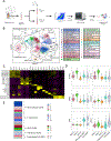
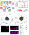
Figure 1.. Multiomic analysis of human carotid atherosclerosis identifies 25 distinct cell populations.
(A) Experimental design. (B) UMAP visualization of clustering revealed 25 cell populations from 88,093 cells across 21 individuals. (C) Canonical Proteins to identify macrophages, T cells, NK cells, B Cells, Plasma Cells, Mast Cells, Neutrophils, dendritic cells, and endothelial cells. (D) Canonical Genes to identify SMCs and Fibroblasts. (E) Distribution of each major subclass across all samples.
Figure 2.
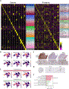
Figure 2.. Multimodal biomarkers of cells within carotid atherosclerotic plaques.
(A) Heatmap of all cell types identified in carotid atherosclerosis. Markers include top 10 mRNA (Left) and top 10 protein (Right) features identified by differential expression. (B) Feature plots displaying unique protein markers for SMCs (Top) and fibroblasts (bottom). (C) Representative immunohistochemistry images from n=5 samples of CD29, CD142, and CD90 in carotid atherosclerosis plaques in early (top row) and late (bottom row) lesions. Scale bars represent 100uM. (D) Feature plots displaying proteins to differentiate endothelial cell 1 and endothelial cell 2. (E) Pathway analysis comparing endothelial cell 1 and endothelial 2 using genes from differential expression analysis.
Figure 3.
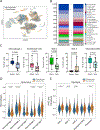
Figure 3.. Cerebrovascular events are associated with alterations in the distribution of certain cell populations.
(A) UMAP colored by clinical status. (B) Vertical bar graph showing proportion of all clusters by clinical status. (C) Box Plots showing distribution of select cell populations including macrophage 1, endothelial cell 1, SMC 2, SMC 3, and modulated SMC. (D) Gene scoring analysis comparing senescence in macrophage clusters in asymptomatic and symptomatic plaques. (E) Gene scoring analysis comparing glycolysis in SMC and fibroblast clusters in asymptomatic and symptomatic plaques.
Figure 4.
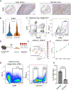
Figure 4.. CD90 expressing cells are associated with advanced atherosclerosis in humans and mice.
(A) Representative immunohistochemistry staining of CD90 in early lesion and necrotic core of same tissue sections. (B) Violin plot comparing CD90 expression in asymptomatic and symptomatic plaques from the CITE-seq analysis. (C) Flow cytometry analysis identifying a subset of cells in human carotid atherosclerotic cell suspensions that express CD90. (D) Experimental design for generation and atherosclerosis induction inof SMC-lineage traced mice. (E) Feature plot displaying the expression of CD90.2 in ZsGreen+ cells in mouse CITE-seq analysis. (F) Proportion of ZsGreen+CD90+ cells during atherosclerosis progression. Line shows mean of n=3. (G) Flow cytometry analysis of aortas from 16-week WD fed LDLr−/−Myh11-CreERT2ROSA26ZsGreen+/− mice that identified a significant proportion of CD90+ cells are SMC-derived. (H) Quantification of percentage of CD90+ cells that are either ZsGreen- or ZsGreen+ in flow analysis. Values are shown as mean±SD, n=3.
Figure 5.
Figure 5.. Sub-clustering analysis of myeloid cells reveals further macrophage phenotypic and functional heterogeneity.
(A) Sub-clustering analysis of myeloid cells. Macrophage 1–5, pDC, and cDC clusters were selected from the initial clustering analysis, revealing 10 clusters. (B) Heatmap showing top upregulated pathways for each macrophage subpopulation based on top DE genes. (C) Heatmap showing top predicted transcriptional regulators for each macrophage subpopulation. (D) UMAP overlaying expression of common M1 and M2 signatures (left), and violin plots quantifying median expression for each macrophage subpopulation. (E) Gene scoring analysis for inflammasome activation, DNA damage, and efferocytosis across macrophage subpopulations.
Figure 6.
Figure 6.. Sub-clustering of SMC and Fibroblasts reveals distinct subpopulations.
(A) Sub-clustering analysis of SMC and fibroblasts. SMC 1–3, modulated SMC, and fibroblast clusters were selected from the initial analysis, revealing 10 clusters. (B) Dotplot showing expression of classic SMC genes with corresponding phenotypes. (C) Radar plot showing pathways for SMCs obtained from DE genes. (D) Radar plot showing pathways for fibroblasts obtained from DE genes. (E and F) Immunohistochemistry staining targeting foamy SMC markers in atherosclerotic tissue sections. (E) carotid artery and (F) coronary artery. DAPI (blue), Smoothelin (green), LipidTOX (red), and CD90 (magenta). White arrows indicate Smoothelin+LipidTOX+CD90+ cells in neointima region of lesion. Colocalization of all 3 markers will appear white. Scale bars=100μm. (G) Box plots showing distribution of select cell populations including foamy SMC, minor SMC 2, and fibromyocyte comparing asymptomatic and symptomatic plaques.

Update of

References

    1. Libby P. The changing landscape of atherosclerosis. Nature. 2021;592:524–533. doi: 10.1038/s41586-021-03392-8 - DOI - PubMed
    1. Bentzon JF, Otsuka F, Virmani R, Falk E. Mechanisms of plaque formation and rupture. Circ Res. 2014;114:1852–1866. doi: 10.1161/CIRCRESAHA.114.302721 - DOI - PubMed
    1. Burke AP, Farb A, Malcom GT, Liang YH, Smialek J, Virmani R. Coronary risk factors and plaque morphology in men with coronary disease who died suddenly. N Engl J Med. 1997;336:1276–1282. doi: 10.1056/NEJM199705013361802 - DOI - PubMed
    1. Finn AV, Nakano M, Narula J, Kolodgie FD, Virmani R. Concept of vulnerable/unstable plaque. Arterioscler Thromb Vasc Biol. 2010;30:1282–1292. doi: 10.1161/ATVBAHA.108.179739 - DOI - PubMed
    1. Aragam KG, Jiang T, Goel A, et al. Discovery and systematic characterization of risk variants and genes for coronary artery disease in over a million participants. Nat Genet. 2022;54:1803–1815. doi: 10.1038/s41588-022-01233-6 - DOI - PMC - PubMed

MeSH terms