Review
. 2024 Mar 14;120(3):223-236.
doi: 10.1093/cvr/cvae021.

Endothelial to mesenchymal transition: at the axis of cardiovascular health and disease

Affiliations
Review

Endothelial to mesenchymal transition: at the axis of cardiovascular health and disease

Ignacio Fernando Hall et al. Cardiovasc Res. .

Abstract

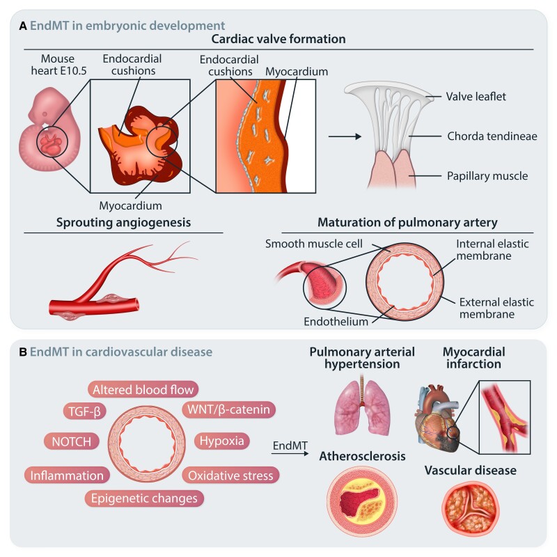
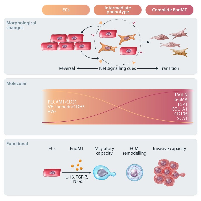
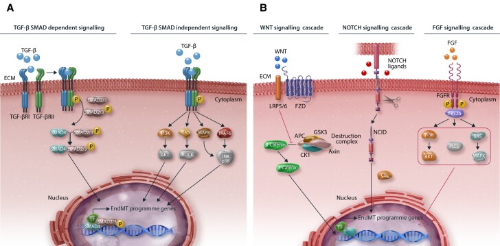
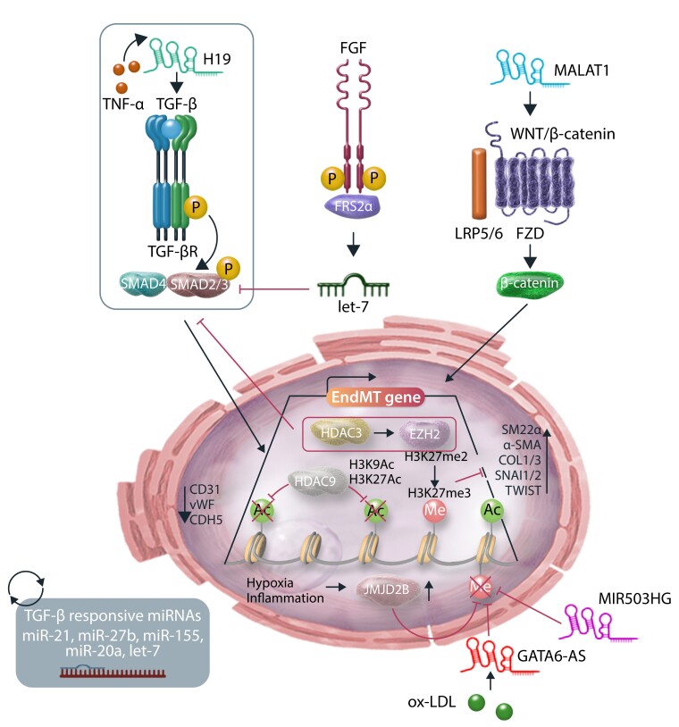
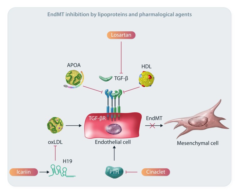
Endothelial cells (ECs) line the luminal surface of blood vessels and play a major role in vascular (patho)-physiology by acting as a barrier, sensing circulating factors and intrinsic/extrinsic signals. ECs have the capacity to undergo endothelial-to-mesenchymal transition (EndMT), a complex differentiation process with key roles both during embryonic development and in adulthood. EndMT can contribute to EC activation and dysfunctional alterations associated with maladaptive tissue responses in human disease. During EndMT, ECs progressively undergo changes leading to expression of mesenchymal markers while repressing EC lineage-specific traits. This phenotypic and functional switch is considered to largely exist in a continuum, being characterized by a gradation of transitioning stages. In this report, we discuss process plasticity and potential reversibility and the hypothesis that different EndMT-derived cell populations may play a different role in disease progression or resolution. In addition, we review advancements in the EndMT field, current technical challenges, as well as therapeutic options and opportunities in the context of cardiovascular biology.

Keywords: Cellular plasticity and heterogeneity; Development; Endothelial cell biology; Endothelial-to-mesenchymal transition (EndMT); Human disease; Therapeutic options.


Conflict of interest statement

Conflict of interest: none declared.

Figures

Figure 1
Figure 1
EndMT in health and disease. (A) Schematic representation of EndMT in embryonic development. A subset of ECs lining the AV canal undergo EndMT and mediate endocardial cushion formation. Further endocardial cushion remodelling mediates healthy AV valve formation. Developmental EndMT is also reported in sprouting angiogenesis and in the maturation of pulmonary arteries. (B) In adult life, EndMT has been associated with the onset and progression of numerous cardiovascular diseases and certain malignancies.
Figure 2
Figure 2
Overview of the EndMT process. Graphical summary of the complex morphological, molecular, and functional changes characterizing EndMT. In response to different stimuli (such as IL-1β, TGF-β, and TNF-α), ECs are activated and undergo EndMT to differentiate towards mesenchymal-like cells. Morphologically, ECs gradually lose their cobblestone structure and cell–cell junctions to acquire an elongated phenotype. This is accompanied by reduced EC-specific marker expression (e.g. CD31, CDH5, and vWF) and increased mesenchymal markers (e.g. TAGLN, α-SMA, FSP-1, COL1A1, CD105, and SCA1). ECs may undergo intermediate or complete EndMT based on net signalling cues. Intermediate EndMT gives rise to intermediary cells that coexpress endothelial and mesenchymal markers. Ultimately, EndMT-derived mesenchymal and mesenchymal-like cells lose most of their endothelial functions and show increased migratory and invasive capacities.
Figure 3
Figure 3
Key signalling pathways involved in EndMT modulation. (A) Graphical illustration of TGF-β signalling (B) and other common pathways in EndMT. For SMAD-dependent signalling, TGF-β binds to and activates the TGF-β I/II receptor complex. This results in the recruitment of SMAD2/3 proteins, which form a complex together with SMAD4. This is translocated into the nucleus to induce the expression of EndMT-associated genes. SMAD-independent TGF-β signalling pathways include, among others, MAPK, RHO, PI3K, and TRAF6. Other common signalling cascades that regulate EndMT include WNT, NOTCH, and FGF. Binding of WNT to the LRP5/6-FZD receptor complex mediates translocation of β-catenin into the nucleus by promoting de-assembly of the β-catenin destruction complex (APC-GSK3-Axin-CK1). NOTCH signalling activation involves the cleavage, release, and translocation of NICD into the nucleus and subsequent EndMT induction. FGF signalling involves the induction of downstream PI3K, PLCγ, and RAS signalling cascades mediated by FRS2α anchoring to FGFR and subsequent EndMT inhibition.
Figure 4
Figure 4
Epigenetic mechanisms and regulation of EndMT. Schematic representation of epigenetic modifications and ncRNA-mediated regulation of EndMT. HDAC3 recruits EZH2 that via H3K27me3 deposition silences TGF-β signalling and blocks EndMT. HDAC9 induces EndMT by repressing H3K9 and H3K27 acetylation. Histone demethylase JMJD2B advances EndMT by demethylating the repressive H3K9me3 at promoters of EndMT controlling genes. LncRNAs H19 and MALAT1 activate the EndMT transcriptional programme by activating the TGF-β and WNT/β-catenin cascades, respectively. LncRNAs GATA6-AS and MIR503HG act by inhibiting EndMT and maintaining EC identity. Many miRNAs have been implicated in regulating EndMT, by either inhibiting or promoting this process, either directly or indirectly. For example, dysregulation of FGF signalling results in reduced let-7 miRNA levels, which in turn increases TGF-β signalling and induces EndMT.
Figure 5
Figure 5
Possible therapeutic avenues of EndMT modulation. Mechanistic details of representative EndMT inhibitors and their associated pathways.

References

    1. Petty RG, Pearson JD. Endothelium--the axis of vascular health and disease. J R Coll Physicians Lond 1989;23:92–102. - PMC - PubMed
    1. Cines DB, Pollak ES, Buck CA, Loscalzo J, Zimmerman GA, McEver RP, Pober JS, Wick TM, Konkle BA, Schwartz BS, Barnathan ES, McCrae KR, Hug BA, Schmidt AM, Stern DM. Endothelial cells in physiology and in the pathophysiology of vascular disorders. Blood 1998;91:3527–3561. - PubMed
    1. Atkins GB, Jain MK, Hamik A. Endothelial differentiation: molecular mechanisms of specification and heterogeneity. Arterioscler Thromb Vasc Biol 2011;31:1476–1484. - PMC - PubMed
    1. Potente M, Mäkinen T. Vascular heterogeneity and specialization in development and disease. Nat Rev Mol Cell Biol 2017;18:477–494. - PubMed
    1. Eelen G, de Zeeuw P, Treps L, Harjes U, Wong BW, Carmeliet P. Endothelial cell metabolism. Physiol Rev 2018;98:3–58. - PMC - PubMed

Publication types