. 2024 May;40(5):422-434.
doi: 10.1002/kjm2.12813. Epub 2024 Feb 22.

GLI family zinc finger protein 2 promotes skin fibroblast proliferation and DNA damage repair by targeting the miR-200/ataxia telangiectasia mutated axis in diabetic wound healing

Affiliations

GLI family zinc finger protein 2 promotes skin fibroblast proliferation and DNA damage repair by targeting the miR-200/ataxia telangiectasia mutated axis in diabetic wound healing

Zun-Hong Liang et al. Kaohsiung J Med Sci. 2024 May.

Abstract

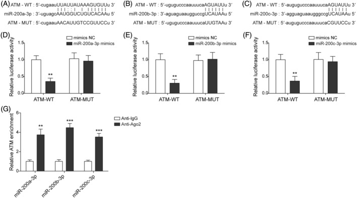
Diabetic foot ulcer (DFU) is a serious complication of diabetic patients which negatively affects their foot health. This study aimed to estimate the role and mechanism of the miR-200 family in DNA damage of diabetic wound healing. Human foreskin fibroblasts (HFF-1 cells) were stimulated with high glucose (HG). Db/db mice were utilized to conduct the DFU in vivo model. Cell viability was evaluated using 3-(4,5-dimethyl-2-thiazolyl)-2,5-diphenyl-2-H-tetrazolium bromide assays. Superoxide dismutase activity was determined using detection kits. Reactive oxygen species determination was conducted via dichlorodihydrofluorescein-diacetate assays. Enzyme-linked immunosorbent assay was used to evaluate 8-oxo-7,8-dihydro-2'deoxyguanosine levels. Genes and protein expression were analyzed by quantitative real-time polymerase chain reaction, western blotting, or immunohistochemical analyses. Luciferase reporter gene and RNA immunoprecipitation assays determined the interaction with miR-200a/b/c-3p and GLI family zinc finger protein 2 (GLI2) or ataxia telangiectasia mutated (ATM) kinase. HG repressed cell proliferation and DNA damage repair, promoted miR-200a/b/c-3p expression, and suppressed ATM and GLI2. MiR-200a/b/c-3p inhibition ameliorated HG-induced cell proliferation and DNA damage repair repression. MiR-200a/b/c-3p targeted ATM. Then, the silenced ATM reversed the miR-200a/b/c-3p inhibition-mediated alleviative effects under HG. Next, GLI2 overexpression alleviated the HG-induced cell proliferation and DNA damage repair inhibition via miR-200a/b/c-3p. MiR-200a/b/c-3p inhibition significantly promoted DNA damage repair and wound healing in DFU mice. GLI2 promoted cell proliferation and DNA damage repair by regulating the miR-200/ATM axis to enhance diabetic wound healing in DFU.

Keywords: ATM; DFU; DNA damage repair; GLI2; miR‐200 family.


Conflict of interest statement

The authors declare that they have no conflict of interest.

Figures

FIGURE 1
FIGURE 1
High glucose (HG) repressed cell proliferation and DNA damage repair in human foreskin fibroblasts (HFF‐1 cells). HFF‐1 cells were stimulated with normal glucose (NG), mannitol (MA), and HG. (A) 3‐(4,5‐dimethyl‐2‐thiazolyl)‐2,5‐diphenyl‐2‐H‐tetrazolium bromide assays estimated cell viability. (B) Dichlorodihydrofluorescein‐diacetate assays determined ROS levels (Scale bar = 100 μm). (C) Enzyme‐linked immunosorbent assay assays evaluated 8‐oxo‐7,8‐dihydro‐2′deoxyguanosine (8‐oxo‐dG) levels. (D) Superoxide dismutase (SOD) activity was measured using SOD detection assays. (E) Quantitative real‐time polymerase chain reaction detected the relative expression of miR‐200a/b/c‐3p. (F). Western blot detected the expression of GLI family zinc finger protein 2 (GLI2) and ataxia telangiectasia mutated (ATM). Each experiment was independently repeated three times. *p < 0.05, **p < 0.01, ***p < 0.001.
FIGURE 2
FIGURE 2
The MiR‐200 inhibitor alleviated the high glucose (HG)‐induced repression of DNA damage repair. Human foreskin fibroblasts were treated with HG and transfected with the miR‐200a/b/c‐3p inhibitor and inhibitor negative control (NC). (A) Quantitative real‐time polymerase chain reaction (qRT‐PCR) detected the expression of miR‐200a/b/c‐3p under normal glucose (NG) conditions. (B) qRT‐PCR detected the relative expression of miR‐200a/b/c‐3p under HG conditions. (C) 3‐(4,5‐dimethyl‐2‐thiazolyl)‐2,5‐diphenyl‐2‐H‐tetrazolium bromide assays estimated cell viability. (D) Dichlorodihydrofluorescein‐diacetate assays determined the ROS levels (Scale bar = 100 μm). (E) Enzyme‐linked immunosorbent assay (ELISA) assays evaluated 8‐oxo‐7,8‐dihydro‐2′deoxyguanosine (8‐oxo‐dG) levels. (F) Superoxide dismutase (SOD) activity was measured by SOD detection assays. (G). Western blot detected ataxia telangiectasia mutated (ATM) expression. Each experiment was independently repeated three times. *p < 0.05, **p < 0.01, ***p < 0.001.
FIGURE 3
FIGURE 3
MiR‐200 targeted ataxia telangiectasia mutated (ATM) and downregulated its expression. Human foreskin fibroblasts were transfected with miR‐200a/b/c‐3p mimics and mimics NC. (A–C) Bioinformatics predicted the binding sites between ATM and miR‐200a/b/c‐3p. (D–F) Dual‐luciferase reporter assays were used to determine the interaction between ATM and miR‐200a/b/c‐3p. (G) The interaction between ATM and miR‐200a/b/c‐3p was validated by RNA immunoprecipitation assays. Each experiment was independently repeated three times. *p < 0.05, **p < 0.01, ***p < 0.001.
FIGURE 4
FIGURE 4
Ataxia telangiectasia mutated (ATM) silencing reversed the regulation of the miR‐200 inhibitor in DNA damage repair under HG conditions. Human foreskin fibroblasts were treated with high glucose (HG) and transfected with the miR‐200a/b/c‐3p inhibitor, inhibitor negative control (NC), si‐ATM, and si‐NC. (A) Quantitative real‐time polymerase chain reaction assays the detected ATM expression. (B) 3‐(4,5‐dimethyl‐2‐thiazolyl)‐2,5‐diphenyl‐2‐H‐tetrazolium bromide assays the estimated cell viability. (C) Dichlorodihydrofluorescein‐diacetate assays determined the reactive oxygen species levels (Scale bar = 100 μm). (D) Enzyme‐linked immunosorbent assay assays evaluated 8‐oxo‐7,8‐dihydro‐2′deoxyguanosine (8‐oxo‐dG) levels. (E) Superoxide dismutase (SOD) activity was measured by SOD detection assays. Each experiment was independently repeated three times. *p < 0.05, **p < 0.01, ***p < 0.001. DMSO, dimethyl sulfoxide; GLI2, GLI family zinc finger protein 2.
FIGURE 5
FIGURE 5
Overexpressed GLI family zinc finger protein 2 (GLI2) ameliorated the high glucose (HG)‐induced repression of DNA damage repair through the modulation of miR‐200. Human foreskin fibroblasts were treated with HG and transfected with pcDNA3.1‐GLI2, pcDNA3.1‐NC, miR‐200a/b/c‐3p mimics, and mimics negative control (NC). (A) Dual‐luciferase reporter assays determined the interaction between GLI2 and miR‐200a/b/c‐3p. (B) Quantitative real‐time polymerase chain reaction (qRT‐PCR) was used to detect the expression of miR‐200a/b/c‐3p. (C) Western blot checked GLI2 expression. (D) qRT‐PCR detected the expression of miR‐200a/b/c‐3p. (E) 3‐(4,5‐dimethyl‐2‐thiazolyl)‐2,5‐diphenyl‐2‐H‐tetrazolium bromide assays estimated cell viability. (F) Dichlorodihydrofluorescein‐diacetate assays determined ROS levels (Scale bar = 100 μm). (G) Enzyme‐linked immunosorbent assay assays evaluated 8‐oxo‐7,8‐dihydro‐2′deoxyguanosine (8‐oxo‐dG) levels. (H) Superoxide dismutase (SOD) activity was measured by SOD detection assays. (I). Western blot detected the protein expression of ataxia telangiectasia mutated (ATM). Each experiment was independently repeated three times. *p < 0.05, **p < 0.01, ***p < 0.001.
FIGURE 6
FIGURE 6
MiR‐200 inhibition promoted foot wound healing by enhancing DNA damage repair in the diabetic foot ulcer (DFU) mice model. DFU mice were administrated with antagomiR‐200a/b/c, antago‐negative control (NC), and liraglutide treatment. (A) Quantitative real‐time polymerase chain reaction detected the miR‐200a/b/c‐3p expression. (B) Wound healing in DFU mice was recorded on days 0, 5, 10, and 15. (C) Western blot examined the expression of ataxia telangiectasia mutated (ATM). (D) Immunohistochemical analysis assessed ATM expression (Scale bar = 100 μm). Each experiment was independently repeated three times. *p < 0.05, **p < 0.01, ***p < 0.001.

References

    1. Fard AS, Esmaelzadeh M, Larijani B. Assessment and treatment of diabetic foot ulcer. Int J Clin Pract. 2007;61(11):1931–1938. - PubMed
    1. Reardon R, Simring D, Kim B, Mortensen J, Williams D, Leslie A. The diabetic foot ulcer. Aust J Gen Pract. 2020;49(5):250–255. - PubMed
    1. Rai V, Moellmer R, Agrawal DK. Stem cells and angiogenesis: implications and limitations in enhancing chronic diabetic foot ulcer healing. Cell. 2022;11(15):2287. - PMC - PubMed
    1. Spampinato SF, Caruso GI, De Pasquale R, Sortino MA, Merlo S. The treatment of impaired wound healing in diabetes: looking among old drugs. Pharmaceuticals (Basel). 2020;13(4):60. - PMC - PubMed
    1. Liang ZH, Pan YC, Lin SS, Qiu ZY, Zhang Z. LncRNA MALAT1 promotes wound healing via regulating miR‐141‐3p/ZNF217 axis. Regen Ther. 2020;15:202–209. - PMC - PubMed