. 2024 Jul 5;147(7):2471-2482.
doi: 10.1093/brain/awae058.

ZSCAN10 deficiency causes a neurodevelopmental disorder with characteristic oto-facial malformations

Affiliations

ZSCAN10 deficiency causes a neurodevelopmental disorder with characteristic oto-facial malformations

Lucia Laugwitz et al. Brain. .

Abstract

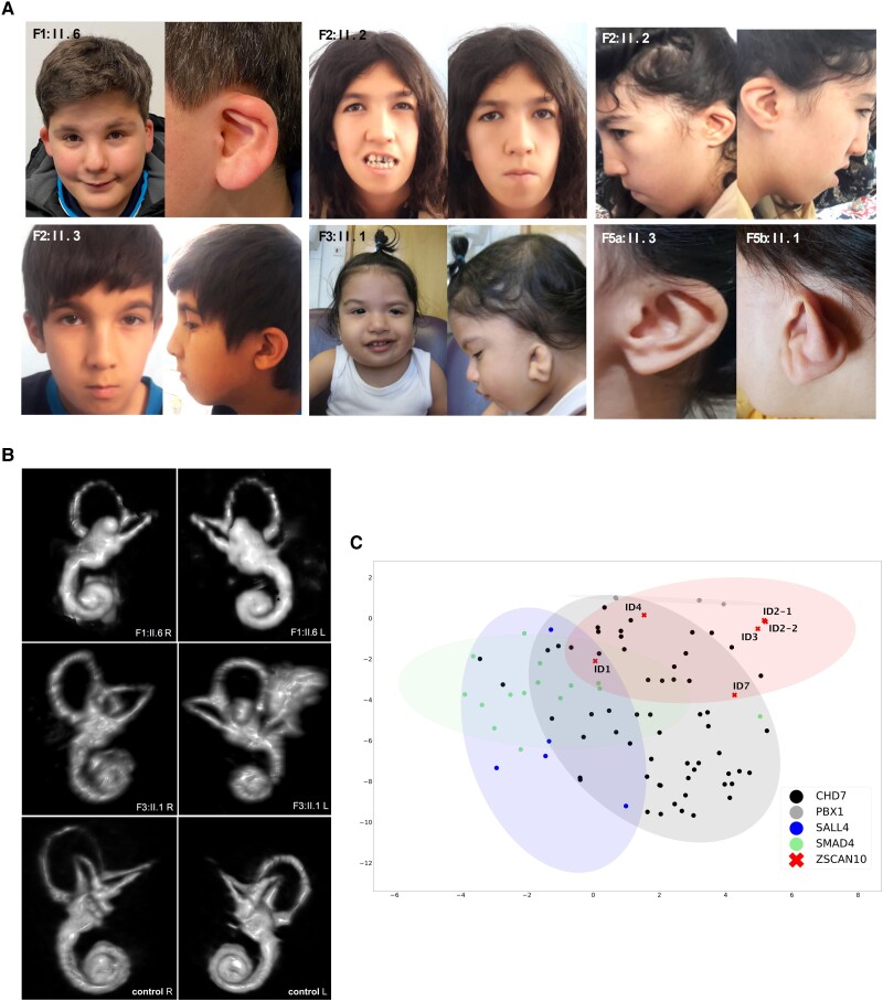
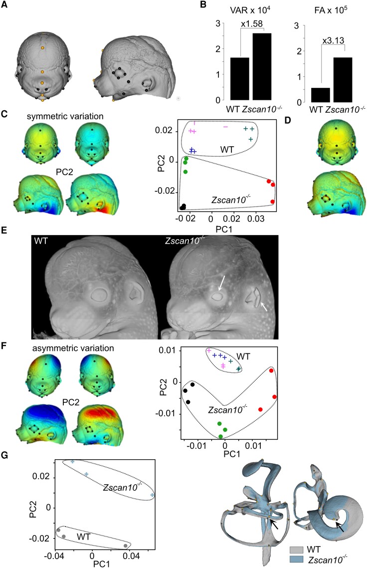
Neurodevelopmental disorders are major indications for genetic referral and have been linked to more than 1500 loci including genes encoding transcriptional regulators. The dysfunction of transcription factors often results in characteristic syndromic presentations; however, at least half of these patients lack a genetic diagnosis. The implementation of machine learning approaches has the potential to aid in the identification of new disease genes and delineate associated phenotypes. Next generation sequencing was performed in seven affected individuals with neurodevelopmental delay and dysmorphic features. Clinical characterization included reanalysis of available neuroimaging datasets and 2D portrait image analysis with GestaltMatcher. The functional consequences of ZSCAN10 loss were modelled in mouse embryonic stem cells (mESCs), including a knockout and a representative ZSCAN10 protein truncating variant. These models were characterized by gene expression and western blot analyses, chromatin immunoprecipitation and quantitative PCR (ChIP-qPCR) and immunofluorescence staining. Zscan10 knockout mouse embryos were generated and phenotyped. We prioritized bi-allelic ZSCAN10 loss-of-function variants in seven affected individuals from five unrelated families as the underlying molecular cause. RNA-sequencing analyses in Zscan10-/- mESCs indicated dysregulation of genes related to stem cell pluripotency. In addition, we established in mESCs the loss-of-function mechanism for a representative human ZSCAN10 protein truncating variant by showing alteration of its expression levels and subcellular localization, interfering with its binding to DNA enhancer targets. Deep phenotyping revealed global developmental delay, facial asymmetry and malformations of the outer ear as consistent clinical features. Cerebral MRI showed dysplasia of the semicircular canals as an anatomical correlate of sensorineural hearing loss. Facial asymmetry was confirmed as a clinical feature by GestaltMatcher and was recapitulated in the Zscan10 mouse model along with inner and outer ear malformations. Our findings provide evidence of a novel syndromic neurodevelopmental disorder caused by bi-allelic loss-of-function variants in ZSCAN10.

Keywords: neurodevelopmental disorders; oto-facial syndrome; semicircular canal dysplasia; zinc finger transcription factor.


Conflict of interest statement

B.B. is Co-Founder and Shareholder of AIRAmed GmbH. The other authors report no competing interests.

Figures

Figure 1
Figure 1
Pedigrees of investigated families and structure of ZSCAN10. (A) Pedigrees of five families with pathogenic variants in ZSCAN10, illustrating affected (filled symbol), healthy (open symbol) family members. Heterozygous carriers are indicated (open symbols containing central dot). Unaffected siblings and fetuses were not tested unless indicated. Families F5a and F5b were remotely related. Individual F5b:II.1 exhibited thalassaemia minor as an additional phenotype. Individual F5b.II.2 was intentionally aborted due to genetically confirmed thalassaemia major (asterisk); the fetus was a carrier of the c.1456C>T variant in ZSCAN10. (B) Structure of ZSCAN10 and the encoded protein with known domains and position of identified variants. aa = amino acid; CDS = coding sequence.
Figure 2
Figure 2
Characterization of Zscan10 in mouse endothelial stem cells and its functional impact. (A) Top: Schematic diagram showing the strategy to knock out the Zscan10 gene in mouse endothelial stem cells (mESCs). Two gRNAs targeting upstream (gRNA1) and downstream (gRNA2) of Zscan10 exon 2 were used to knock out exon 2 of the Zscan10 gene in mESCs. Bottom: RNA-sequencing (RNA-seq) data coverage plot showing the expression of the Zscan10 gene in wild-type and Zscan10 knockout mESCs (Zscan10−/−). Exon 2 of Zscan10 is lost in Zscan10−/− cell lines (grey shaded area). (B) Volcano plot showing the differentially expressed genes (DEGs) in Zscan10−/− mESCs compared with control mESCs, including 710 downregulated genes and 600 upregulated genes. The genes regulated by Zscan10 and related to pluripotency and differentiation of ESCs are labelled. FC = fold-change; FDR = false discovery rate. (C) Top: Schematic diagrams showing the structures of wild-type ZSCAN10 (ZSCAN10 full) and ZSCAN10 c.1456C>T variant (expressing only the first 485 amino acids of the ZSCAN10 protein) expression vectors. Bottom: Representative western blot showing expression of ZSCAN10 full and ZSCAN10 485 protein. (D) Chromatin immunoprecipitation and quantitative PCR (ChIP-qPCR) shows the binding affinity of ZSCAN10 full, ZSCAN10 485 mutated protein on the Pou5f1 promoter in different vector transfected mESCs. Empty vector transfected mESCs were used as a negative control. ****P < 0.0001. (E) RNA-seq data coverage plot showing the expression of the Pou5f1 gene in wild-type and Zscan10−/− mESCs. Chr17 = chromosome 17. (F) Representative images of immunofluorescence staining show the subcellular distribution of ZSCAN10 full and ZSCAN10 485 proteins. The wild-type ZSCAN10 protein (ZSCAN10 full) is located mainly in the nucleus, whereas the mutant ZSCAN10 protein (ZSCAN10 485) is located mainly in the cytoplasm.
Figure 3
Figure 3
Facial dysmorphisms and neuroimaging findings in subjects with ZSCAN10 deficiency. (A) Dysmorphic features in ZSCAN10 deficiency include facial asymmetry (Individual F1:II.6 and F3:II.1) with reduced facial movements, outer ear malformations, down slanting palpebral fissures and prominent epicanthic folds. (B) MRI-based 3D reconstruction of the inner ear. Right (R) and left (L) inner ear reconstructions showing bilateral aplasia of the horizontal semicircular canals (SCC) and dysplasia of the vestibule in Individual F1:II.6 and SCC dysplasia in Individual F3:II.1 as well as a healthy individual as the control. (C) 2D visualization of 90 images from individuals with pathogenic variants in ZSCAN10, CHD7, PBX1, SALL4 and SMAD4 by t-distributed stochastic neighbour embedding.
Figure 4
Figure 4
Facial dysmorphism in Zscan10-deficient mouse embryos. (A) Twenty-six surface landmarks (10 paired in black plus six unpaired in orange) were affixed in triplicates on three independent wild-type (WT) and three Zscan10−/− embryonic Day (E)14.5 embryos. (B) Among-individual Procrustes variance and fluctuating asymmetry (FA). Variance (VAR) estimates were multiplied by 104 and FA by 105. (C) Principal components of symmetric variation. Left: Coloured meshes correspond to variation (negative–positive) along PC1 (left) or PC2 (right). Blue corresponds to contraction and red to expansion compared with the opposite side, i.e. the shape on left corner corresponds to the shape inferred for the negative PC2 and indicates the deviation from the shape on its right; for instance, in the knockout (KO) there is a relative opening of the ear compared with the WT. Right: Colours correspond to individuals (three replicates per individual): filled circles represent KO individuals and crosses represent WT individuals. (D) Differences between symmetric averages. Shape corresponds to the least-squared (LS) means of the KO and the colours show the expansion or contraction (blue) from the WT. Shape changes are very similar to PC2 (α = 18°) from C. (E) Representative head volumes for WT and Zscan10−/− E14.5 embryos showing smaller eye size and ear opening (white arrows). (F) Principal components of asymmetric variation indicate that Zscan10-deficient and WT mice appear to have very different asymmetry patterns, with Zscan10-deficient mice displaying larger and more fluctuating deviations. (G) Principal components of inner ear reconstitution. Average reconstitutions were computed and overlayed (grey for WT and blue for Zscan10−/−) showing a mild difference between Zscan10-deficient and WT mice with PC2 accounting for 20% of the variance corresponding to a misalignment of one of the semicircular tubes and a shortening of the cochlea (black arrow).

References

    1. Sheridan E, Wright J, Small N, et al. Risk factors for congenital anomaly in a multiethnic birth cohort: An analysis of the Born in Bradford study. Lancet. 2013;382:1350–1359. - PubMed
    1. Maulik PK, Mascarenhas MN, Mathers CD, Dua T, Saxena S. Prevalence of intellectual disability: A meta-analysis of population-based studies. Res Dev Disabil. 2011;32:419–436. - PubMed
    1. Kochinke K, Zweier C, Nijhof B, et al. Systematic phenomics analysis deconvolutes genes mutated in intellectual disability into biologically coherent modules. Am J Hum Genet. 2016;98:149–164. - PMC - PubMed
    1. Deciphering Developmental Disorders Study . Prevalence and architecture of de novo mutations in developmental disorders. Nature. 2017;542:433–438. - PMC - PubMed
    1. Kaplanis J, Samocha KE, Wiel L, et al. Evidence for 28 genetic disorders discovered by combining healthcare and research data. Nature. 2020;586:757–762. - PMC - PubMed