. 2024 Feb 27;121(9):e2320657121.
doi: 10.1073/pnas.2320657121. Epub 2024 Feb 22.

Inverse regulation of SOS1 and HKT1 protein localization and stability by SOS3/CBL4 in Arabidopsis thaliana

Affiliations

Inverse regulation of SOS1 and HKT1 protein localization and stability by SOS3/CBL4 in Arabidopsis thaliana

Francisco Gámez-Arjona et al. Proc Natl Acad Sci U S A. .

Abstract

To control net sodium (Na+) uptake, Arabidopsis plants utilize the plasma membrane (PM) Na+/H+ antiporter SOS1 to achieve Na+ efflux at the root and Na+ loading into the xylem, and the channel-like HKT1;1 protein that mediates the reverse flux of Na+ unloading off the xylem. Together, these opposing transport systems govern the partition of Na+ within the plant yet they must be finely co-regulated to prevent a futile cycle of xylem loading and unloading. Here, we show that the Arabidopsis SOS3 protein acts as the molecular switch governing these Na+ fluxes by favoring the recruitment of SOS1 to the PM and its subsequent activation by the SOS2/SOS3 kinase complex under salt stress, while commanding HKT1;1 protein degradation upon acute sodic stress. SOS3 achieves this role by direct and SOS2-independent binding to previously unrecognized functional domains of SOS1 and HKT1;1. These results indicate that roots first retain moderate amounts of salts to facilitate osmoregulation, yet when sodicity exceeds a set point, SOS3-dependent HKT1;1 degradation switches the balance toward Na+ export out of the root. Thus, SOS3 functionally links and co-regulates the two major Na+ transport systems operating in vascular plants controlling plant tolerance to salinity.

Keywords: Arabidopsis; HKT1; SOS pathway; salinity; sodium transport.


Conflict of interest statement

Competing interests statement:The authors declare no competing interest.

Figures
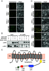
Fig. 1.
Fig. 1.
SOS3 interacts with SOS1. (A) SOS1-SOS3 interaction visualized by BiFC. SOS1 and SOS2 fused to the N terminus of the fluorescent protein YFP, and wild-type SOS3 and mutant SOS3-1 proteins fused to the C terminus of YFP, were transiently co-expressed in N. benthamiana leaves in the combinations indicated. Combinations that included an empty vector produced no fluorescence. SOS1 fused to full-length GFP (lower row) was used as a control to label the PM. Fluorescence signals for interaction were detected by confocal microscopy 2 to 3 d after infiltration. (Scale bar is 20 μm.) (B) Co-immunoprecipitation of SOS1 and SOS3. The translational fusion SOS1:GFP was transiently co-expressed with MYC-tagged wild-type SOS3 (SOS3:MYC) or the mutant protein SOS3-1 (SOS3-1:MYC) in N. benthamiana leaves. Total proteins were extracted and SOS1:GFP and associating proteins were pulled down with α-GFP antibody. SDS-PAGE and immunoblots were performed with α-GFP and α-MYC antibodies to detect the proteins expressed (Input) and pulled-down (IP). (C) Schematic representation of SOS1 topology and known functional domains. The N-terminal transmembrane segments (gray) extend up to amino acid Gly-441. The large cytosolic part of SOS1 contains the cyclic-nucleotide binding homologous domain (blue, G765-L841), the auto-inhibitory domain (red, L1005-L1047), and the phosphorylation site by the SOS2/SOS3 kinase complex at Ser-1138 (green). The SOS3 binding domain (K460-L482) is shown in yellow. Amino acid residues flanking each domain are indicated. (D) The SOS3 binding domain of SOS1 is necessary and sufficient for interaction. Full-length SOS1, or with an internal deletion of the SOS3 binding domain (SOS1ΔS3BD), or the SOS3 binding domain alone (S3BD) were fused to the N-terminal part of YFP and co-expressed in N. benthamiana leaves with SOS3 fused to the C-terminal part of YFP. The C-terminally truncated SOS1 protein at Gln-998 that lacks the AID and SOS2 phosphorylation site (SOS1Δ998) was fused to the N terminus of YFP, whereas SOS2 was fused to the C terminus of YFP. Recombinant proteins were co-expressed as indicated in each row. Fluorescence signals were detected by confocal microscopy 3 d after infiltration. (Scale bar is 20 μm.)
Fig. 2.
Fig. 2.
SOS3 binding to SOS1. (A) Fluorescence spectroscopy titration of SOS3 with S3BD. The squares represent the average value of three independent series of experiments, whose individual values are shown as dots. The error bars are SD. (B) Predicted structure of the S3BD of SOS1. Residues forming the hydrophobic surface exposed to the solvent are indicated. (C) Complex of SOS3 bound to the S3BD of SOS1. The hydrophobic surface of S3BD faces the crevice of SOS3 that serves to bind interacting proteins. Residue W177 used to analyze protein binding is indicated. (D) SOS3 bound to the SOS1 dimer. The SOS3-S3BD complex (C) was overlaid with the structure of the SOS1 dimer. The dashed line separates the transmembrane domain forming the transporting pore of SOS1 and the helical domain underneath the membrane, which contains the S3BD.
Fig. 3.
Fig. 3.
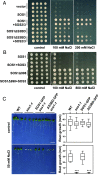
Relevance of SOS3 binding for SOS1 activity. (A) Full-length SOS1 or a recombinant protein in which the SOS3-binding domain had been spliced out (SOS1ΔS3BD) was expressed alone or together with SOS2 and SOS3 in yeast cells to reconstitute a functional SOS module. Four serial decimal dilutions (10-1 to 10-4) of overnight liquid cultures of transformed cells were spotted in selective AP medium supplemented with the indicated NaCl concentrations. Plates were pictured after 2 to 3 d at 30 °C. Two independent transformants for each combination are shown. (B) Full-length SOS1 or a constitutively active SOS1 protein lacking the auto-inhibitory and phosphorylation domains (SOS1Δ998) was co-expressed or not with SOS3. Samples were processed as in (A). Note that maximal NaCl concentration used was 800 mM. (C) Mutant sos1-1 seedlings transformed with constructs pSOS1:SOS1:GFP or pSOS1:SOS1ΔS3BD:GFP to express wild-type SOS1 or a recombinant form with the SOS3 binding domain spliced out (SOS1ΔS3BD) at nearly native protein levels, were initially grown on regular ½MS medium and then transferred to fresh media plates with and without 50 mM NaCl to test for phenotypic complementation. The box plots show root growth. Centerlines show the medians; box limits indicate the 25th and 75th percentiles; whiskers extend to the minimum and maximum values (n = 14). Asterisks indicate significantly different means relative to WT, based on one-way ANOVA followed by Tukey’s multiple comparisons test, P < 0.001.
Fig. 4.
Fig. 4.
Salinity- and SOS3-dependent recruitment of SOS1 to the PM. (A) Representative spinning disk confocal images of epidermal cells at the root meristem of Col-0 gl1 (wild-type, WT) and sos3-1 transgenic lines expressing pSOS1:SOS1:GFP and treated with 50 mM NaCl over 1 to 3 d. (Scale bar is 10 μm.) (B) Quantification of SOS1:GFP fluorescence partition between PM and intracellular compartments (IC) compartments of samples shown in (A). Violin plots: centerlines show the medians, dotted lines indicate the 25th and 75th percentiles. Data are from n ≥ 70 cells from ≥7 plants. Letters indicate significantly different means, One-way ANOVA followed by Tukey’s HSD, P < 0.05. (C) Representative spinning disk confocal images of epidermal cells at the root meristem of sos1-1 plants transformed with the indicated plasmids. Samples were shortly treated with FM4-64 to stain the PM and imaged in control conditions. (Scale bars, 10 μm.) (D) Quantification of fluorescence partition between PM and IC compartments of samples in (C). Violin plots are as in (B). Data are from 60 cells of 6 plants. Asterisks indicate significantly different means at P < 0.0001, Welch’s unpaired t test.
Fig. 5.
Fig. 5.
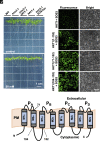
Physical and genetic interactions of SOS3 and HKT1. (A) Mutation hkt1-1 suppresses the salt hypersensitivity of sos3-1. Seedlings of wild-type, mutants sos3-1, hkt1-1, hkt1-1 sos3-1, and the complemented line sos3-1 35S:SOS3 (SOS3ox) were transferred to ½MS medium supplemented with 50 and 100 mM NaCl. Pictures were taken 1 wk after transfer. (Scale bar size is 1 cm.) (B) BiFC assay to map the interaction domain of HKT1;1 with SOS3. SOS3 fused to the C-terminal part of the Venus fluorescent protein (SOS3:VC) was transiently co-expressed in tobacco epidermal cells with the Venus N-terminal part fused to the full-length HKT1;1 (HKT1:VN) or the indicated protein fragments. Fluorescence signals produced by protein interaction were detected by confocal microscopy on the third day after infiltration. (Scale bar size is 20 μm.) (C) Schematic representation of HKT1;1 protein topology consisting of 4 pore-loop domains (PA to PD) and the transmembrane fragments (M1A/M2A to M1D/M2D).
Fig. 6.
Fig. 6.
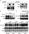
SOS3 controls HKT1 protein stability. (A) The HKT1:GFP fusion protein was transiently co-expressed with SOS3:MYC or SOS3-1:MYC in tobacco leaves. Total proteins were extracted and HKT1:GFP-associating proteins were pulled down with α-GFP antibody. SDS-PAGE and immunoblots were performed to detect expressed proteins (Input) and pulled-down proteins (IP). α-GFP and α-MYC antibodies were used to detect HKT1;1 and SOS3 or SOS3-1, respectively. (B) Total proteins were extracted from 2-wk-old Arabidopsis wild-type seedlings transformed with 35S:HKT1:GFP and treated with 100 mM NaCl for the indicated periods. The HKT1:GFP protein was detected by western blot using α-GFP antibody. Coomassie Brilliant Blue (CBB) staining is shown as a loading reference. (C) Arabidopsis seedlings of genotype hkt1-3 transformed with pHKT1:HKT1:CFP levels were salt-treated 100 mM NaCl, with and without 50 µM MG132, for 12 h. The HKT1:CFP protein was detected with α-GFP antibody, and α-tubulin antibodies were used for protein loading control. (D) Ten-day-old seedlings of wild-type, sos1-1, sos2-2, and sos3-1 transformed with construct 35S:HKT1:GFP were treated with 100 mM NaCl and with or without 100 µM MG132 for 12 h, and total protein was extracted from root tissues. Immunoblots were performed to detect HKT1:GFP protein with α-GFP. CBB staining and western blotting of HSP90 show protein loading.
Fig. 7.
Fig. 7.
SOS3 controls long-distance Na+ transport from roots to shoots. (A) Na+ concentration in roots and leaves. Two-week old plants of genotypes Col-0 gl1 (WT), hkt1-1, sos1-1, sos3-1, and the double mutant sos1-1 sos3-1 were transferred to hydroponic LAK medium supplemented with 5 mM NaCl and 0.1 mM KCl for another 2 wk. Plants were collected, weighted, and had their root and leaves separated and homogenized individually. Shown are the means and SE of the Na+ contents (% of dry weight) determined by atomic emission spectrometry. The ratios of root-to-shoot contents are given in the Lower panel. Different letters indicate statistical differences by Fisher’s LSD (P < 0.05). Smaller values are given in numerals for clarity. (B) Mechanistic model of SOS3 operation. Under non-saline or moderately saline conditions, roots take up salts, including Na+ ions, to reduce root water potential and draw water from the soil. The net balance of basal activity of SOS1 and the retrieval of Na+ ions from the xylem sap by HKT1;1 system allows Na+ uptake and the retention of salts in the root. When salinity reaches stress levels, activated SOS3 promotes the delivery of SOS1 to the PM and enhances its Na+/H+ exchanger activity via phosphorylation by SOS2. Whether binding of SOS3 to SOS1 and SOS1 activation by the SOS2/SOS3 complex (not shown) occurs simultaneously or they proceed stepwise so that SOS1 is first trafficked to the PM and subsequently activated by SOS2/SOS3, remains unknown. In concert, activated SOS3 targets HKT1;1 for degradation to prevent further Na+ unloading from the xylem sap. These events lead to increased Na+ efflux toward the soil and active loading into the xylem, thereby allowing the evapotranspiration stream to evacuate Na+ toward the shoot, where potentially toxic Na+ ions will be compartmentalized into vacuoles and diluted by growth.

References

    1. Deinlein U., et al. , Plant salt-tolerance mechanisms. Trends Plant Sci. 19, 371–379 (2014). - PMC - PubMed
    1. Cheeseman J. M., The evolution of halophytes, glycophytes and crops, and its implications for food security under saline conditions. New Phytol. 206, 557–570 (2015). - PubMed
    1. Ismail A. M., Horie T., Genomics, physiology, and molecular breeding approaches for improving salt tolerance. Annu. Rev. Plant Biol. 68, 405–434 (2017). - PubMed
    1. Ji H., et al. , The Salt Overly Sensitive (SOS) pathway: Established and emerging roles. Mol. Plant 6, 275–286 (2013). - PubMed
    1. El Mahi H., et al. , A critical role of sodium flux via the plasma membrane Na(+)/H(+) exchanger SOS1 in the salt tolerance of rice. Plant Physiol. 180, 1046–1065 (2019). - PMC - PubMed

Substances

LinkOut - more resources