. 2024 Mar 7;111(3):529-543.
doi: 10.1016/j.ajhg.2024.02.001. Epub 2024 Feb 21.

De novo variants in DENND5B cause a neurodevelopmental disorder

Collaborators, Affiliations

De novo variants in DENND5B cause a neurodevelopmental disorder

Marcello Scala et al. Am J Hum Genet. .

Abstract

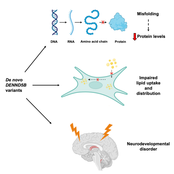
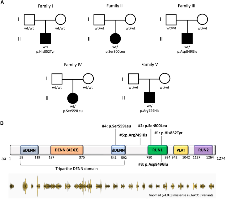
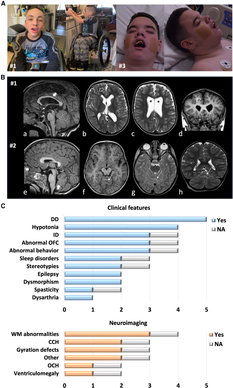
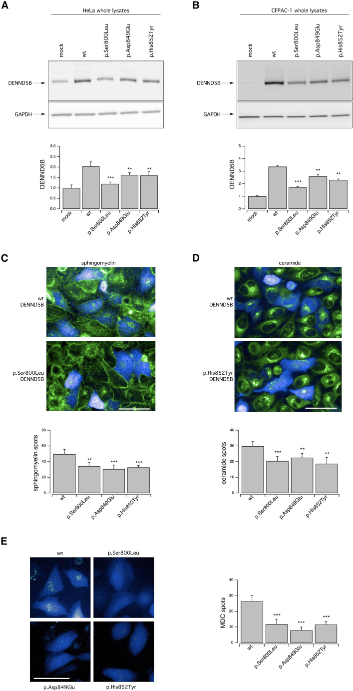
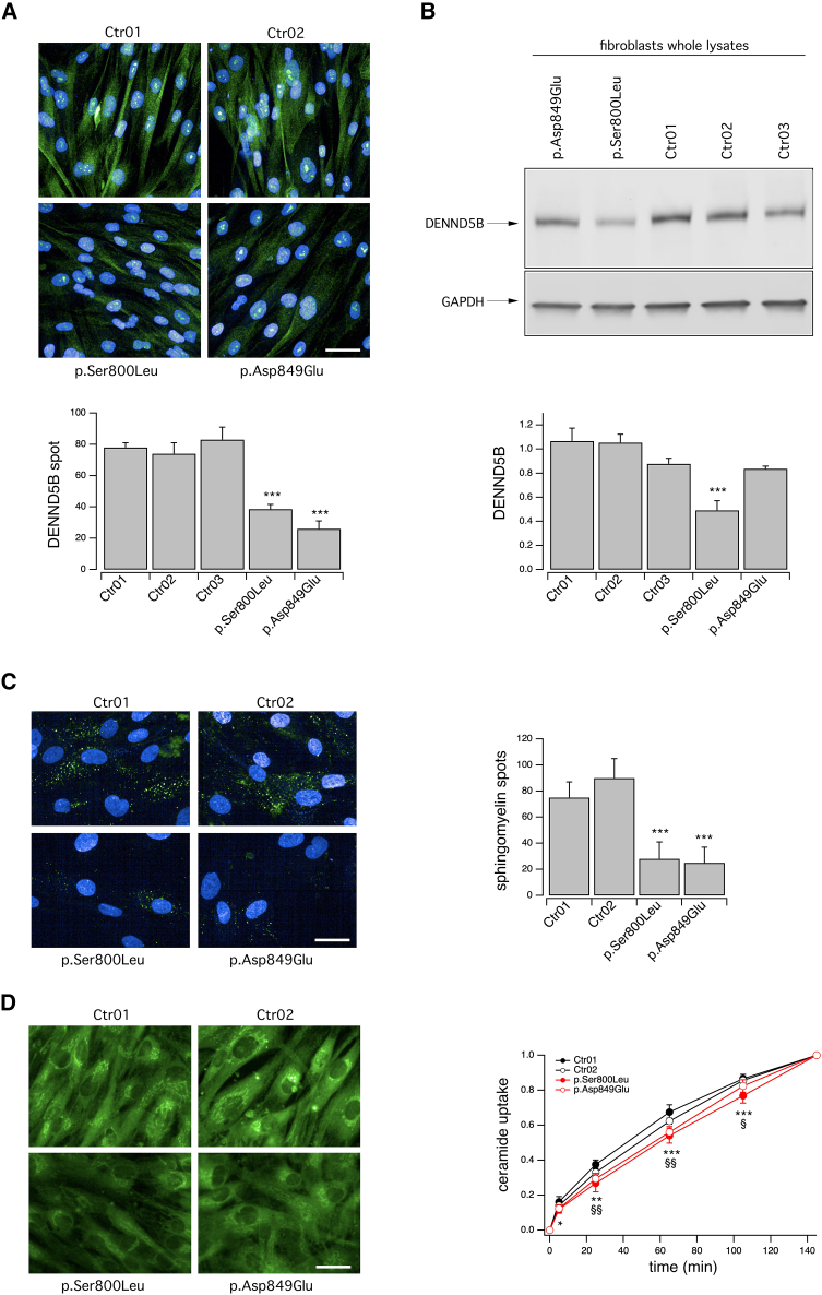
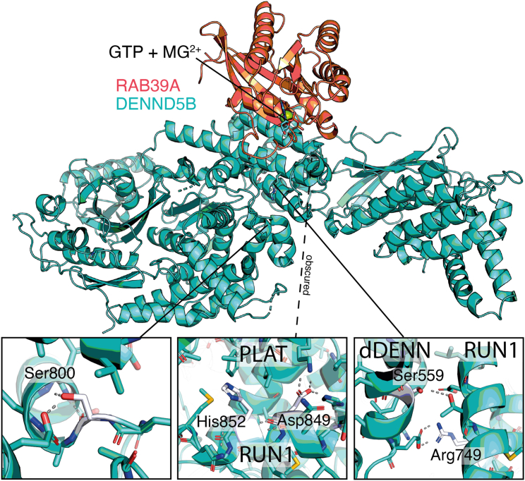
The Rab family of guanosine triphosphatases (GTPases) includes key regulators of intracellular transport and membrane trafficking targeting specific steps in exocytic, endocytic, and recycling pathways. DENND5B (Rab6-interacting Protein 1B-like protein, R6IP1B) is the longest isoform of DENND5, an evolutionarily conserved DENN domain-containing guanine nucleotide exchange factor (GEF) that is highly expressed in the brain. Through exome sequencing and international matchmaking platforms, we identified five de novo variants in DENND5B in a cohort of five unrelated individuals with neurodevelopmental phenotypes featuring cognitive impairment, dysmorphism, abnormal behavior, variable epilepsy, white matter abnormalities, and cortical gyration defects. We used biochemical assays and confocal microscopy to assess the impact of DENND5B variants on protein accumulation and distribution. Then, exploiting fluorescent lipid cargoes coupled to high-content imaging and analysis in living cells, we investigated whether DENND5B variants affected the dynamics of vesicle-mediated intracellular transport of specific cargoes. We further generated an in silico model to investigate the consequences of DENND5B variants on the DENND5B-RAB39A interaction. Biochemical analysis showed decreased protein levels of DENND5B mutants in various cell types. Functional investigation of DENND5B variants revealed defective intracellular vesicle trafficking, with significant impairment of lipid uptake and distribution. Although none of the variants affected the DENND5B-RAB39A interface, all were predicted to disrupt protein folding. Overall, our findings indicate that DENND5B variants perturb intracellular membrane trafficking pathways and cause a complex neurodevelopmental syndrome with variable epilepsy and white matter involvement.

Keywords: DENND5B; Rab GPTases; cell homeostasis; epilepsy; guanine nucleotide exchange factors; intellectual disability; lipid uptake and distribution; membrane trafficking; neurodevelopmental disorder.


Conflict of interest statement

Declaration of interests A.C. is an employee of GeneDx, LLC. The Department of Molecular and Human Genetics at Baylor College of Medicine receives revenue from clinical genetic testing completed at Baylor Genetics Laboratories.

Figures

None
Graphical abstract
Figure 1
Figure 1
Summary of DENND5B variants and family pedigrees (A) Schematic representation of DENND5B (GenBank: NP_659410.3) showing the localization of the variants detected in the reported cohort. Most variants lie within or in close proximity to the tripartite DENN domain or the RUN1 domain. (B) Pedigrees of the reported families showing the segregation of DENND5B variants (GenBank: NM_144973.4). Abbreviations: DENN, differentially expressed in neoplastic vs. normal cells; NA, not available; PLAT, polycystin-1, lipoxygenase, alpha-toxin; RUN, RPIP8 (RaP2-interacting protein 8), UNC-14, and NESCA.
Figure 2
Figure 2
Clinical and neuroimaging details of subjects harboring DENND5B variants (A) Clinical photographs of subjects harboring DENND5B variants. Subject #1 shows macrocephaly, mild intellectual disability, axial hypotonia, and appendicular spasticity. Facial dysmorphism includes arched and sparse eyebrows, narrow palpebral fissures, hypertelorism, bulbous nose tip, deep nasal bridge, thick lower lip, and prominent chin. Subject #3 has severe cognitive deficiency, microcephaly, and hypotonia. Dysmorphic features include short nose, hypertelorism, malposition of teeth, deep nasal bridge, deep philtrum, and micrognathia. (B) Neuroimaging findings. (a–d) Brain MRI of subject #1 with sagittal T1-weighted (a), axial T2-weighted (b, c), and coronal T1-weighted (d) showing a thin corpus callosum (thick arrow), a small anterior commissure (thin arrow), white matter volume loss with ventricular enlargement (asterisks), and a focal gyral anomaly with infolding in the right frontal lobe (empty arrows). (e–h) Brain MRI of subject #2 with sagittal T1-weighted (a), axial T1-weighted (b), axial T2-weighted (c), and coronal T2-weighted (d) revealing focal foliar anomaly in the superior cerebellar vermis (dashed arrows) associated with inferior vermis hypoplasia (thick arrow). (C) Histogram graphs showing the distribution of clinical features and brain MRI abnormalities in the cohort of subjects harboring DENND5B variants. Abbreviations: CCH, corpus callosum hypoplasia; DD, developmental delay; ID, intellectual disability; NA, not available; OCH, optic chiasm hypoplasia; OFC, occipito-frontal circumference; WM, white matter.
Figure 3
Figure 3
Biochemical and functional analysis of WT and mutant DENND5B proteins in heterologous expression systems DENND5B proteins were transiently expressed in different cell models by means of vectors encoding the different DENND5B proteins and a TagBFP2-based expression marker that allowed easy monitoring of transfection efficiency. (A and B) Western blot analysis (top) and densitometric quantification (bottom) of the levels of the WT, p.Ser800Leu, p.Asp849Glu, and p.His852Tyr DENND5B proteins in whole lysates of HeLa (A) and CFPAC-1 (B) cells. Lysates of mock-transfected parental cells have been included to show the expression level of the endogenous DENND5B protein in the corresponding cell model. Data are means ± SEM (n = 3). (C and D) Representative confocal microscopy images (top) of C12-sphingomyelin (C) or C6-ceramide (D) uptake and quantification (bottom) of fluorescent spots resembling intracellular vesicles containing the corresponding lipid probe. The uptake was assayed on HeLa cells following transient expression of the WT, p.Ser800Leu, p.Asp849Glu, and p.His852Tyr DENND5B constructs. Quantification of spots was performed on TagBFP2-positive cells. Data are means ± SEM (n = 5). For each independent biological replicate, a region measuring 1,320 × 1,320 μm was imaged and analyzed, comprising approximately 35–40 TagBFP2-positive cells. (E) Representative confocal microscopy images (left) and quantification (right) of fluorescent spots resembling autophagic vacuoles visualized using the MDC probe on HeLa cells following transient expression of the WT, p.Ser800Leu, p.Asp849Glu, and p.His852Tyr DENND5B constructs. Quantification of spots was performed on TagBFP2-positive cells. Data are means ± SEM (n = 5). For each independent biological replicate, a region measuring 1,320 × 1,320 μm was imaged and analyzed, comprising approximately 35–40 TagBFP2-positive cells. Asterisks indicate statistical significance vs. control (WT-DENND5B-transfected cells): ∗∗p < 0.01; ∗∗∗p < 0.001. Scale bar = 100 μm.
Figure 4
Figure 4
Biochemical and functional characterization of DENND5B variants in affected subjects’ fibroblasts (A) Representative confocal microscopy images (top) and quantification (bottom) of DENND5B-positive spots following immunolocalization of DENND5B protein in fibroblasts derived from subjects #2 and #3, carrying, respectively, the p.Ser800Leu and p.Asp849Glu variant and, for comparison, fibroblasts derived from non-NDD subjects. (B) Western blot analysis (top) and densitometric quantification (bottom) of the DENND5B levels in whole lysates of fibroblasts derived from affected individuals and control subjects. Data are means ± SEM (n = 5). (C) Representative confocal microscopy images (left) of C12-sphingomyelin uptake and quantification (right) of fluorescent spots resembling intracellular vesicles containing the lipid probe. The uptake was assayed on fibroblasts derived from affected individuals and control subjects. Data are means ± SEM (n = 9). (D) Representative confocal microscopy images (left) of C6-ceramide uptake and intracellular distribution and its quantification (right). The uptake was assayed on fibroblasts derived from affected individuals and control subjects. Data are means ± SEM (n = 9). For each independent biological replicate, a region measuring 990 × 1,320 μm was imaged and analyzed, comprising approximately one hundred fibroblasts. Asterisks indicate statistical significance: p < 0.05; ∗∗p < 0.01; ∗∗∗p < 0.001 for p.Ser800Leu fibroblasts vs. pool of control fibroblasts; §p < 0.05; §§p < 0.01; for p.Asp849Glu fibroblasts vs. pool of control fibroblasts. Scale bar = 100 μm.
Figure 5
Figure 5
In silico analysis of DENND5B variants Serine 559 (mutated to leucine in #4) is localized in the dDENN domain. This is a surface mutation facing and in close proximity to the RUN domain, within 5 Å of threonine 776, which is the length of a water bridge. Arginine 749 (histidine 749 in #5) is in the 1st RUN domain. This is a surface variant facing the dDENN domain and act as potentially pathogenic via the same mechanism as p.Ser559Leu. Serine 800 (leucine 800 in #2) is in the 1st RUN domain. This is the last residue in a turn and forms a hydrogen bond with glutamate 786 in the previous helix via its side chain, which would be lost in a leucine variant. In fact, predictions suggest it to be highly destabilizing (>10 kcal/mol). Aspartate 849 (glutamate 849 in #3) is localized in the 1st RUN domain. This buried charged residue forms salt bridges with lysine 876 and histine 882. Despite the similarity of the two residues, glutamate is longer and predictions suggest it to be highly destabilizing (6 kcal/mol). Histidine 852 (tyrosine 852 in #1) is localized in the 1st RUN domain. This buried residue is on the following rung of the helix as aspartate 849 and its side chain appears packed in two alternative conformations depending on the distance to β-hairpin 1015–1019 of the PLAT/LH2 (which shifts by 1 Å). Both conformations form several hydrophobic interactions, but in the more distant conformation (shown), it forms π-π interactions with tryptophan 927 and a π-sulfur interaction with methionine 856, while in the other it forms a hydrogen bond with the backbone of a preceding residue. If this actually represents two biologically relevant states, any change would disrupt this balance.

References

    1. Marat A.L., Dokainish H., McPherson P.S. DENN domain proteins: regulators of Rab GTPases. J. Biol. Chem. 2011;286:13791–13800. - PMC - PubMed
    1. Yoshimura S.i., Gerondopoulos A., Linford A., Rigden D.J., Barr F.A. Family-wide characterization of the DENN domain Rab GDP-GTP exchange factors. J. Cell Biol. 2010;191:367–381. - PMC - PubMed
    1. Homma Y., Hiragi S., Fukuda M. Rab family of small GTPases: an updated view on their regulation and functions. FEBS J. 2021;288:36–55. - PMC - PubMed
    1. Zerial M., McBride H. Rab proteins as membrane organizers. Nat. Rev. Mol. Cell Biol. 2001;2:107–117. - PubMed
    1. Mignogna M.L., D’Adamo P. Critical importance of RAB proteins for synaptic function. Small GTPases. 2018;9:145–157. - PMC - PubMed

Publication types

MeSH terms