. 2024 Jul 18;64(1):2301612.
doi: 10.1183/13993003.01612-2023. Print 2024 Jul.

Long-term radiological and pulmonary function abnormalities at 3 years after COVID-19 hospitalisation: a longitudinal cohort study

Affiliations

Long-term radiological and pulmonary function abnormalities at 3 years after COVID-19 hospitalisation: a longitudinal cohort study

Xiaoyu Han et al. Eur Respir J. .

Abstract

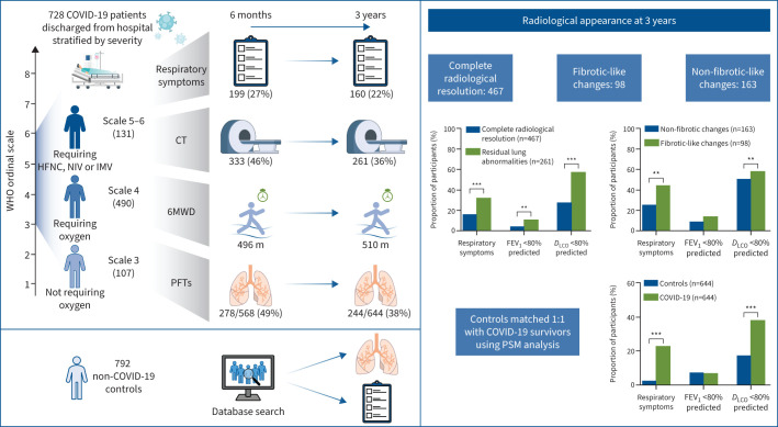
Background: This study aimed to evaluate the longitudinal progression of residual lung abnormalities (ground-glass opacities, reticulation and fibrotic-like changes) and pulmonary function at 3 years following coronavirus disease 2019 (COVID-19).

Methods: This prospective, longitudinal cohort study enrolled COVID-19 survivors who exhibited residual lung abnormalities upon discharge from two hospitals. Follow-up assessments were conducted at 6 months, 12 months, 2 years and 3 years post-discharge, and included pulmonary function tests, 6-min walk distance (6MWD), chest computed tomography (CT) scans and symptom questionnaires. Non-COVID-19 controls were retrospectively recruited for comparative analysis.

Results: 728 COVID-19 survivors and 792 controls were included. From 6 months to 3 years, there was a gradual improvement in reduced diffusing capacity of the lung for carbon monoxide (D LCO <80% predicted: 49% versus 38%; p=0.001), 6MWD (496 versus 510 m; p=0.002) and residual lung abnormalities (46% versus 36%; p<0.001), regardless of disease severity. Patients with residual lung abnormalities at 3 years more commonly had respiratory symptoms (32% versus 16%; p<0.001), lower 6MWD (494 versus 510 m; p=0.003) and abnormal D LCO (57% versus 27%; p<0.001) compared with those with complete resolution. Compared with controls, the proportions of D LCO impairment (38% versus 17%; p<0.001) and respiratory symptoms (23% versus 2.2%; p<0.001) were significantly higher in the matched COVID-19 survivors at the 3-year follow-up.

Conclusions: Most patients exhibited improvement in radiological abnormalities and pulmonary function over time following COVID-19. However, more than a third continued to have persistent lung abnormalities at the 3-year mark, which were associated with respiratory symptoms and reduced diffusion capacity.


Conflict of interest statement

Conflict of interest: The authors have no potential conflicts of interest to disclose.

Figures

None
Summary of the main study findings. Severity scale 3: patients not requiring supplemental oxygen. Severity scale 4: patients requiring supplemental oxygen via nasal cannula or mask. Severity scale 5–6: patients requiring high-flow nasal cannula (HFNC), non-invasive ventilation (NIV) or invasive mechanical ventilation (IMV) for oxygen therapy. COVID-19: coronavirus disease 2019; WHO: World Health Organization; CT: computed tomography; 6MWD: 6-min walk distance; PFT: pulmonary function test; FEV1: forced expiratory volume in 1 s; DLCO: diffusing capacity of the lung for carbon monoxide; PSM: propensity score matching. **: p<0.01; ***: p<0.001.
FIGURE 1
FIGURE 1
Study flowcharts. a) Flow diagram of coronavirus disease 2019 (COVID-19) patients. b) Matching process of COVID-19 survivors and non-COVID-19 participants who completed pulmonary function tests (PFTs) at the 3-year follow-up visit (1:1). Severity scale 3: patients not requiring supplemental oxygen during hospitalisation. Severity scale 4: patients requiring supplemental oxygen via nasal cannula or mask during hospitalisation. Severity scale 5–6: patients requiring high-flow nasal cannula, non-invasive mechanical ventilation or invasive mechanical ventilation during hospitalisation. CT: computed tomography; 6MWD: 6-min walk distance; HRCT: high-resolution CT.
FIGURE 2
FIGURE 2
Longitudinal evolution of lung function over time. a) Evolution of lung function in all coronavirus disease 2019 (COVID-19) patients who underwent pulmonary function tests. b–d) Evolution of lung function in COVID-19 patients with different disease severity scales: b) severity scale 3 (patients not requiring supplemental oxygen during hospitalisation), c) severity scale 4 (patients requiring supplemental oxygen via nasal cannula or mask during hospitalisation) and d) severity scale 5–6 (patients requiring high-flow nasal cannula, non-invasive mechanical ventilation or invasive mechanical ventilation during hospitalisation). FEV1: forced expiratory volume in 1 s; DLCO: diffusion capacity for carbon monoxide. *: p<0.05 for the comparison of different time-points.
FIGURE 3
FIGURE 3
Dynamic changes observed in computed tomography (CT) scans over time. a) Mean±sd total CT score for patients stratified by different disease severity scales. b–d) The changes in the proportion of residual lung abnormalities among COVID-19 patients classified by different disease severity scales: b) severity scale 3 (patients not requiring supplemental oxygen during hospitalisation), c) severity scale 4 (patients requiring supplemental oxygen via nasal cannula or mask during hospitalisation) and d) severity scale 5–6 (patients requiring high-flow nasal cannula, non-invasive mechanical ventilation or invasive mechanical ventilation during hospitalisation). *: p<0.05 for the comparison of different time-points.
FIGURE 4
FIGURE 4
Various patterns of computed tomography (CT) evolution in residual lung abnormalities over 3 years. a) Typical findings in participants (n=3) with progressive fibrotic changes. Initial scans revealed extensive bilateral ground-glass opacities (GGOs) with septal thickening (asterisk). The 6-month follow-up displayed focal subpleural reticular lesions in the left upper lobe, evolving into parenchymal bands and cystic lesions at the 12-month follow-up. At 3 years post-symptoms onset, CT scans depicted multiple cystic airspaces and honeycombing (arrows) in the left lower lobe. b) Typical findings in participants (n=95) with stable fibrotic-like changes. Acute-phase scans exhibited diffuse GGOs and consolidation (asterisk) in the right upper and middle lobes. The 6-month follow-up showed subpleural reticular lesions with traction bronchiectasis (arrows), persisting as stable subpleural bronchiectasis in the same lung zone at 12 months and 3 years. c) Typical CT progression in participants (n=75) with complete resolution of non-fibrotic changes. Acute-phase scans displayed GGOs with linear opacities in both lower lobes (asterisks). By the 6-month follow-up, partial resorption was evident (arrows), and by the 3-year follow-up, nearly complete resolution of abnormalities was observed. d) Typical CT findings over time in participants (n=160) with stable non-fibrotic changes. Acute-phase scans revealed a mixed pattern of GGOs and consolidation in the right middle and lower lobes (asterisk). Subsequent scans at 6 months, 2 years and 3 years showed persisting subpleural localised GGOs and reticular lesions in the right lower lobe (arrows).

Comment in

References

    1. World Health Organization . From emergency response to long-term COVID-19 disease management: sustaining gains made during the COVID-19 pandemic. 2023. www.who.int/publications/i/item/WHO-WHE-SPP-2023.1 Date last accessed: 10 February 2024.
    1. Burki T. WHO ends the COVID-19 public health emergency. Lancet Respir Med 2023; 11: 588. doi:10.1016/S2213-2600(23)00217-5 - DOI - PMC - PubMed
    1. Mandal S, Barnett J, Brill SE, et al. . ‘Long-COVID’: a cross-sectional study of persisting symptoms, biomarker and imaging abnormalities following hospitalisation for COVID-19. Thorax 2021; 76: 396–398. doi:10.1136/thoraxjnl-2020-215818 - DOI - PMC - PubMed
    1. Wu X, Liu X, Zhou Y, et al. . 3-month, 6-month, 9-month, and 12-month respiratory outcomes in patients following COVID-19-related hospitalisation: a prospective study. Lancet Respir Med 2021; 9: 747–754. doi:10.1016/S2213-2600(21)00174-0 - DOI - PMC - PubMed
    1. Huang C, Huang L, Wang Y, et al. . 6-month consequences of COVID-19 in patients discharged from hospital: a cohort study. Lancet 2021; 397: 220–232. doi:10.1016/S0140-6736(20)32656-8 - DOI - PMC - PubMed