

Assessment of performance and safety of Corneal Chamber hypothermic storage medium and PSS-L corneal rinsing solution in human and porcine corneas
- PMID: 38388003
- PMCID: PMC10884202
- DOI: 10.1136/bmjophth-2023-001453
Assessment of performance and safety of Corneal Chamber hypothermic storage medium and PSS-L corneal rinsing solution in human and porcine corneas
Abstract
Purpose: To prove the safety and performance of the hypothermic corneal storage medium "Corneal Chamber" and the rinsing solution "PSS-L" in support of the new Conformité Européenne (CE) certification process in accordance with the Medical Device Regulation.
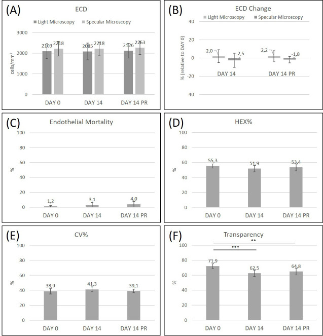
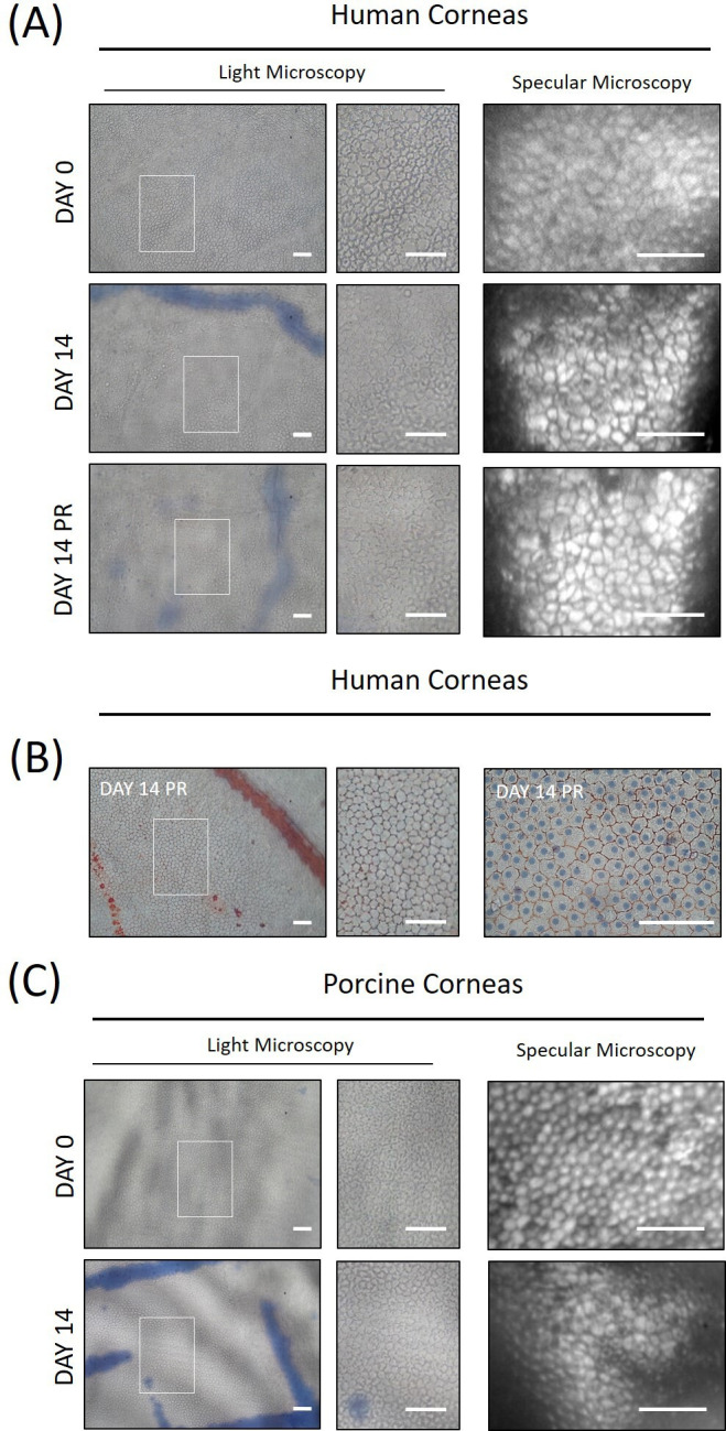
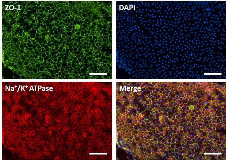
Methods: Fifteen (n=15) human donor corneas and 11 (n=11) porcine corneas were evaluated for the following parameters: endothelial cell density (ECD) and mortality, percentage of hexagonal cells (HEX%), coefficient of cellular area variation (CV%) and corneal transparency at Day 0 and after 14±1 days of storage in Corneal Chamber medium at 2-8°C. Then, the same parameters were assessed after rinsing of corneas in PSS-L for 1 min at room temperature. Evaluation of gentamicin sulfate carryover after corneal storage and PSS-L rinsing was performed by ultra-high performance liquid chromatography analysis on human corneas homogenates.
Results: Human and porcine corneas stored in Corneal Chamber medium showed a good overall quality of the tissue according to the quality parameters evaluated. In particular, mean ECD, HEX% and CV% did not show statistically significant changes at the end of storage and endothelial mortality increased to 3.1±3.3 and 7.8±3.5% in human and porcine corneas, respectively. Tissue rinsing with PSS-L did not affect the quality parameters evaluated before and gentamicin sulfate residues were absent in human corneas.
Conclusions: Corneal preservation in Corneal Chamber medium at 2-8°C for 14 days and the corneal rinse with PSS-L are safe and effective procedures allowing the preservation of the corneal quality parameters as well as the complete elimination of gentamicin sulfate from the tissues before transplantation.Cite Now.
Keywords: Anterior chamber; Cornea; Diagnostic tests/Investigation; Drugs; Experimental & animal models; Experimental & laboratory; Imaging; Treatment Surgery.
© Author(s) (or their employer(s)) 2024. Re-use permitted under CC BY-NC. No commercial re-use. See rights and permissions. Published by BMJ.
Conflict of interest statement
Competing interests: LG, UR, CG, OR and JDT are employees of AL.CHI.MI.A. S.R.L. ER was consulted by AL.CHI.MI.A. S.R.L. for the purpose of conducting statistical analysis.
Figures
Similar articles
-
18 A porcine cornea and lamellar tissue model to investigate effects of storage conditions on corneal preservation.BMJ Open Ophthalmol. 2022 Nov;7(Suppl 2):A8. doi: 10.1136/bmjophth-2022-EEBA.18. BMJ Open Ophthalmol. 2022. PMID: 37282691 Free PMC article.
-
Prospective In Vitro Comparison of Kerasave and Optisol-GS Corneal Storage Solutions.Cornea. 2023 May 1;42(5):630-638. doi: 10.1097/ICO.0000000000003201. Epub 2022 Dec 12. Cornea. 2023. PMID: 36729660
-
Porcine Cornea Storage Ex Vivo Model as an Alternative to Human Donor Tissues for Investigations of Endothelial Layer Preservation.Transl Vis Sci Technol. 2023 Apr 3;12(4):24. doi: 10.1167/tvst.12.4.24. Transl Vis Sci Technol. 2023. PMID: 37079319 Free PMC article.
-
[Transplantation of corneal endothelial cells].Nippon Ganka Gakkai Zasshi. 2002 Dec;106(12):805-35; discussion 836. Nippon Ganka Gakkai Zasshi. 2002. PMID: 12610838 Review. Japanese.
-
Clinical cryobiology of tissues: preservation of corneas.Cryobiology. 1986 Aug;23(4):323-53. doi: 10.1016/0011-2240(86)90038-6. Cryobiology. 1986. PMID: 3527562 Review.
Cited by
-
Retrospective Clinical Outcomes of Keratoplasty Using Human Donor Corneas Preserved in Eusol-C Hypothermic Storage Medium.J Clin Med. 2024 Dec 13;13(24):7606. doi: 10.3390/jcm13247606. J Clin Med. 2024. PMID: 39768528 Free PMC article.
-
A corneal and whole eye globe bovine ex vivo model to mimic human donor corneal storage conditions and eye surgeries.Cell Tissue Bank. 2025 Mar 4;26(2):14. doi: 10.1007/s10561-025-10162-7. Cell Tissue Bank. 2025. PMID: 40038115
References
-
- Macpherson JFE, Souza G, Nicolaissen B, et al. . Comparison of electrolyte composition in four eye bank media during corneal preservation. Int J Eye Bank 2014;2:1–5. 10.7706/ijeb.v2i1.54 - DOI
MeSH terms
Substances
LinkOut - more resources
Full Text Sources