. 2024 Feb 22;14(1):4409.
doi: 10.1038/s41598-024-55050-4.

Polyunsaturated fatty acids-induced ferroptosis suppresses pancreatic cancer growth

Affiliations

Polyunsaturated fatty acids-induced ferroptosis suppresses pancreatic cancer growth

Akane Suda et al. Sci Rep. .

Abstract

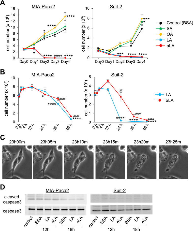
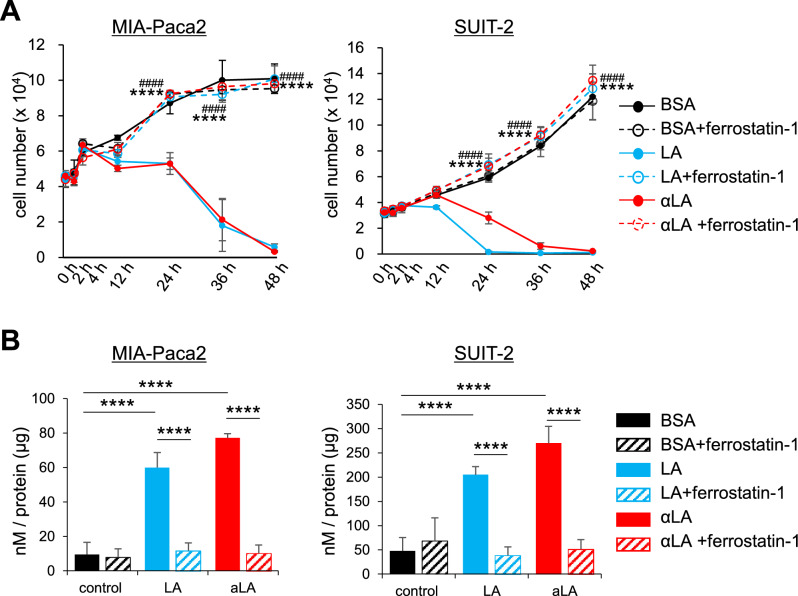
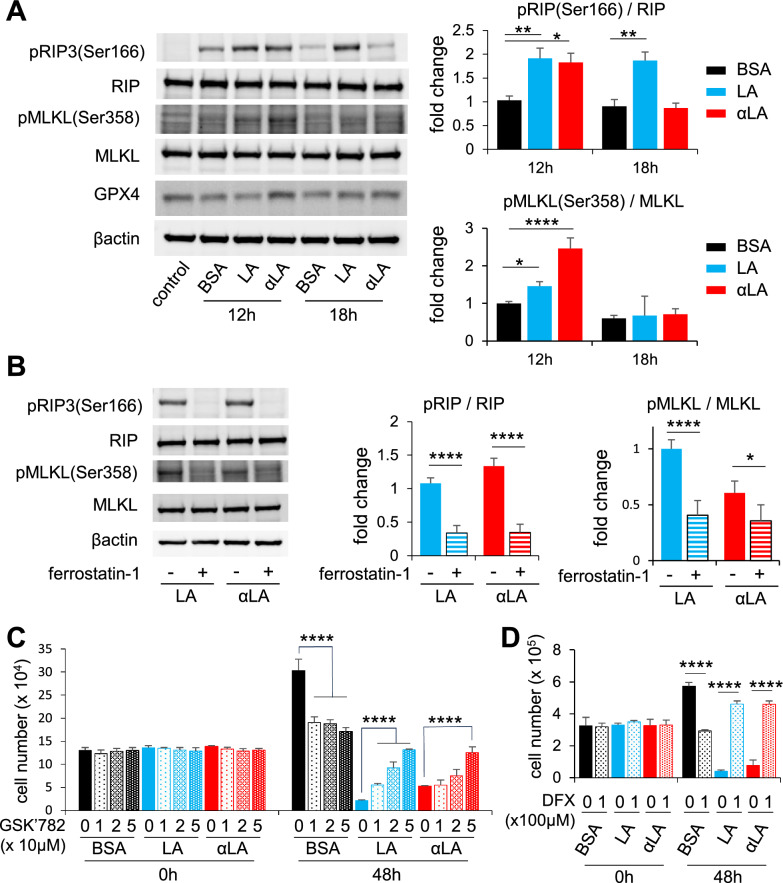
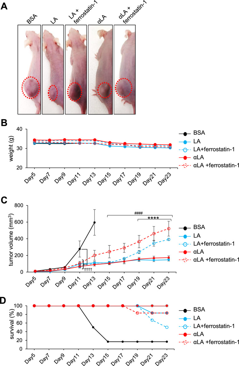
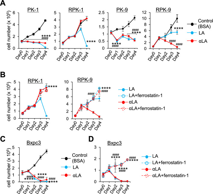
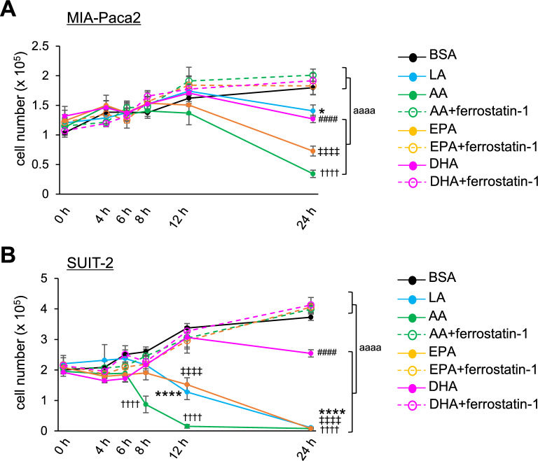
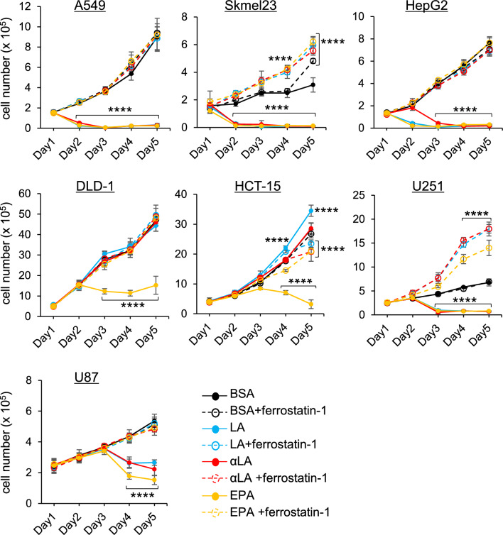
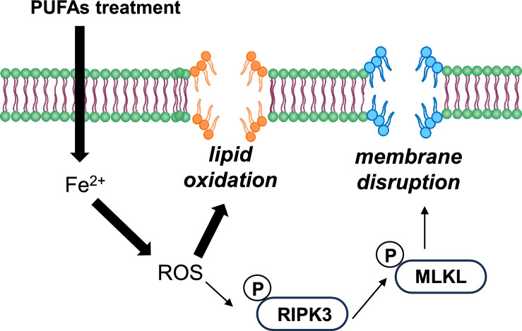
Despite recent advances in science and medical technology, pancreatic cancer remains associated with high mortality rates due to aggressive growth and no early clinical sign as well as the unique resistance to anti-cancer chemotherapy. Current numerous investigations have suggested that ferroptosis, which is a programed cell death driven by lipid oxidation, is an attractive therapeutic in different tumor types including pancreatic cancer. Here, we first demonstrated that linoleic acid (LA) and α-linolenic acid (αLA) induced cell death with necroptotic morphological change in MIA-Paca2 and Suit 2 cell lines. LA and αLA increased lipid peroxidation and phosphorylation of RIP3 and MLKL in pancreatic cancers, which were negated by ferroptosis inhibitor, ferrostatin-1, restoring back to BSA control levels. Similarly, intraperitoneal administration of LA and αLA suppresses the growth of subcutaneously transplanted Suit-2 cells and ameliorated the decreased survival rate of tumor bearing mice, while co-administration of ferrostatin-1 with LA and αLA negated the anti-cancer effect. We also demonstrated that LA and αLA partially showed ferroptotic effects on the gemcitabine-resistant-PK cells, although its effect was exerted late compared to treatment on normal-PK cells. In addition, the trial to validate the importance of double bonds in PUFAs in ferroptosis revealed that AA and EPA had a marked effect of ferroptosis on pancreatic cancer cells, but DHA showed mild suppression of cancer proliferation. Furthermore, treatment in other tumor cell lines revealed different sensitivity of PUFA-induced ferroptosis; e.g., EPA induced a ferroptotic effect on colorectal adenocarcinoma, but LA or αLA did not. Collectively, these data suggest that PUFAs can have a potential to exert an anti-cancer effect via ferroptosis in both normal and gemcitabine-resistant pancreatic cancer.

Keywords: Drug resistance; Ferroptosis; Linoleic acid; Pancreatic cancer; Poly unsaturated fatty acids; α-Linolenic acid.


Conflict of interest statement

The authors declare no competing interests.

Figures

Figure 1
Figure 1
LA and αLA pushed cell death into necroptotic/ferroptotic phenotypes. (A) The proliferation rate of MIA-Paca2 and Suit-2 cells treated with BSA (control) or different 60 µM FAs (SA, OA, LA, and αLA). Live cells were counted successively for 4 days. One-way ANOVA followed by the Tukey test was used for multiple comparisons. Data shown are the means ± SEM *P < 0.05, ***P < 0.001, ****P < 0.0001 versus BSA-control. (B) The proliferation rate of MIA-Paca2 and Suit-2 cells treated with LA, and αLA. Live cells were counted in early time points up to 48 h. Data shown are the means ± SEM *P < 0.05, ****P < 0.0001 versus at 12 h in LA treatment, #P < 0.05, ##P < 0.001, ####P < 0.00001 versus at 12 h in αLA treatment. (C) Representative phase contrast images showing the time course for the morphological change in Mia-Paca2 treated with LA. (D) Western blot for cleaved caspase 3 and caspase 3 in MIA-Paca2 and Suit-2 cells treated with BSA, LA, and αLA for 12 h and 18 h. Control is the cells treated with medium containing 1% FBS only.
Figure 2
Figure 2
LA and αLA induced pancreatic cancer cell death through ferroptosis. (A) The proliferation rate of MIA-Paca2 and Suit-2 cells treated with BSA, 60 µM LA, and 60 µM αLA alone or together with 10 µM ferrostatin-1. Live cells were counted successively up to 48 h. One-way ANOVA followed by the Tukey test was used for multiple comparisons. Data shown are the means ± SEM ****P < 0.00001 versus with LA treatment without ferrostatin-1, ####P < 0.00001 versus with αLA treatment without ferrostatin-1 (B) Bar graph showing lipid peroxidation of LA and αLA in MIA-Paca2 and SUIT-2 cells treated with BSA, 60 µM LA, and 60 µM αLA alone or together with 10 µM ferrostatin-1. Data shown are the means ± SEM ****P < 0.00001.
Figure 3
Figure 3
LA and αLA induced ferroptosis pathway leads to RIP/MLKL dependent necroptosis pathway. (A) Western blot for pRIP3 (Ser166), total RIP, pMLKL (Ser358), total MLKL, and GPX4 in MIA-Paca2 treated with BSA, 60 μM LA, and 60 μM αLA for 12 h and 18 h. Bar graphs show band density of relative phosphorylation levels analyzed using NIH-ImageJ. Data shown are the means ± SEM *P < 0.05, **P < 0.01, ****P < 0.0001 versus BSA-control. (B) Western blot for pRIP3 (Ser166), total RIP, pMLKL (Ser358) and total MLKL in MIA-Paca2 cells treated with 60 µM LA, and 60 µM αLA together with or without 10 µM ferrostatin-1 for 12 h. Bar graphs show band density of relative phosphorylation levels analyzed using NIH-ImageJ. Data shown are the means ± SEM *P < 0.05, ****P < 0.0001 versus BSA-control. (C) The proliferation rate of MIA-Paca2 and Suit-2 cells treated with BSA, 60 µM LA, and 60 µM αLA alone or together with GSK’782 (0, 10, 20, 50 µM). Live cells were counted at 0 h and 48 h. One-way ANOVA followed by the Tukey test was used for multiple comparisons. Data shown are the means ± SEM ****P < 0.00001 versus with LA or αLA treatment without GSK’782. (D) The proliferation rate of MIA-Paca2 and Suit-2 cells treated with BSA, 60 µM LA, and 60 µM αLA alone or together with 100 μM DFX. Live cells were counted at 0 h and 48 h. One-way ANOVA followed by the Tukey test was used for multiple comparisons. Data shown are the means ± SEM ****P < 0.00001 versus with LA or αLA treatment without DFX.
Figure 4
Figure 4
Intraperitoneal administration of LA and αLA suppressed the growth of pancreatic cancer in the xenograft model. (A) Representative images on tumor bearing mice treated with either BSA, 500 mg/kg LA, or 500 mg/kg αLA together with or without 20 mg/kg ferrostatin-1. (B) Average weight of mice treated with BSA as vehicle or 500 mg/kg LA, or 500 mg/kg αLA together with or without 20 mg/kg ferrostatin-1. (C) Cancer growth and (D) corresponding Kaplan–Meier survival curves in mice treated with either BSA, 500 mg/kg LA, or 500 mg/kg αLA together with or without 20 mg/kg ferrostatin-1. One-way ANOVA followed by the Tukey test was used for multiple comparisons. Data shown are the means ± SEM (n = 6 for each group) ****P < 0.00001 between with and without ferrostatin-1 in LA-treated group, ####P < 0.00001 between with and without ferrostatin-1 in αLA-treated group, ┼┼┼┼P < 0.00001 versus BSA-treated group.
Figure 5
Figure 5
LA and αLA had different anti-cancer effects on Gemcitabine-resistant pancreatic cancers. (A and C) The proliferation rate of PK-1, RPK-1, PK-9 and RPK-9 cells (A) and BxPC3 cells (C) treated with BSA, 60 µM LA, and 60 µM αLA (A). Live cells were counted successively for 4 days. One-way ANOVA followed by the Tukey test was used for multiple comparisons. Data shown are the means ± SEM ****P < 0.00001 BSA-control versus LA, ####P < 0.00001 BSA-control and αLA. (B and D) The proliferation rate of RPK-1 and RPK-9 cells (B) and Bxpc3 cells (D) treated with 60 µM LA, and 60 µM αLA alone or together with 10 µM ferrostatin-1. Live cells were counted successively for 4 days. One-way ANOVA followed by the Tukey test was used for multiple comparisons. Data shown are the means ± SEM ****P < 0.00001 between with and without ferrostatin-1 in LA-treated cells, ####P < 0.00001 between with and without ferrostatin-1 in αLA-treated cells.
Figure 6
Figure 6
Ferroptosis effect by PUFA on pancreatic cancers is not dependent on the increasing number of carbon or double bonds in the PUFA. (A and B) The proliferation rate of MIA-Paca2 cells (A) and Suit-2 cells (B) treated with BSA, 60 µM LA, 60 µM AA, 60 µM EPA and 60 µM DHA alone or together with 10 µM ferrostatin-1. Live cells were counted successively up to 24 h. One-way ANOVA followed by the Tukey test was used for multiple comparisons. Data shown are the means ± SEM *P < 0.00001, ####P < 0.00001, ┼┼┼┼P < 0.00001, ǂ ǂ ǂ ǂ P < 0.00001 BSA-control versus LA, DHA, AA, EPA, respectively. aaaaP < 0.00001 between with and without ferrostatin-1 treatment.
Figure 7
Figure 7
Different sensitivity of PUFA-induced ferroptosis. The proliferation rate of A549, Skmel23, HepG2, DLD-1, HCT-15, U251, and U87 cells treated with BSA, 60 µM LA, 60 µM AA, 60 µM αLA, and 60 µM EPA alone or together with 10 µM ferrostatin-1. Live cells were counted successively for 5 days. *P < 0.00001 versus BSA-control.
Figure 8
Figure 8
Schematic illustration how PUFAs induces ferroptotic cell death in the pancreatic cancers. Exogenous PUFAs including LA and αLA taken up into the cells undergo lipid peroxidation leading to ferroptotic cell death. In addition, ROS accumulation from LA and αLA peroxidation may induce phosphorylation of RIP3 and MLKL leading to membrane disruption.

References

    1. Kamisawa T, Wood LD, Itoi T, Takaori K. Pancreatic cancer. Lancet. 2016;388:73–85. doi: 10.1016/S0140-6736(16)00141-0. - DOI - PubMed
    1. Goral V. Pancreatic cancer: Pathogenesis and diagnosis. Asian Pac. J. Cancer Prev. 2015;16:5619–5624. doi: 10.7314/apjcp.2015.16.14.5619. - DOI - PubMed
    1. Grant TJ, Hua K, Singh A. Molecular pathogenesis of pancreatic cancer. Prog. Mol. Biol. Transl. Sci. 2016;144:241–275. doi: 10.1016/bs.pmbts.2016.09.008. - DOI - PMC - PubMed
    1. Gillen S, Schuster T, MeyerZumBuschenfelde C, Friess H, Kleeff J. Preoperative/neoadjuvant therapy in pancreatic cancer: A systematic review and meta-analysis of response and resection percentages. PLoS Med. 2010;7:e1000267. doi: 10.1371/journal.pmed.1000267. - DOI - PMC - PubMed
    1. Suker M, et al. FOLFIRINOX for locally advanced pancreatic cancer: A systematic review and patient-level meta-analysis. Lancet Oncol. 2016;17:801–810. doi: 10.1016/S1470-2045(16)00172-8. - DOI - PMC - PubMed