. 2024 Feb 23;10(1):46.
doi: 10.1186/s40792-024-01843-8.

Ectopic pancreatic adenocarcinoma in Meckel's diverticulum: a case report

Affiliations

Ectopic pancreatic adenocarcinoma in Meckel's diverticulum: a case report

Shoichi Inokuchi et al. Surg Case Rep. .

Abstract

Background: Malignant neoplasms arising from Meckel's diverticulum are rare and an adenocarcinoma in Meckel's diverticulum originating from ectopic pancreatic tissue is even rarer. Herein, we report a patient with an ectopic pancreatic adenocarcinoma in Meckel's diverticulum who was successfully treated with surgery and chemotherapy.

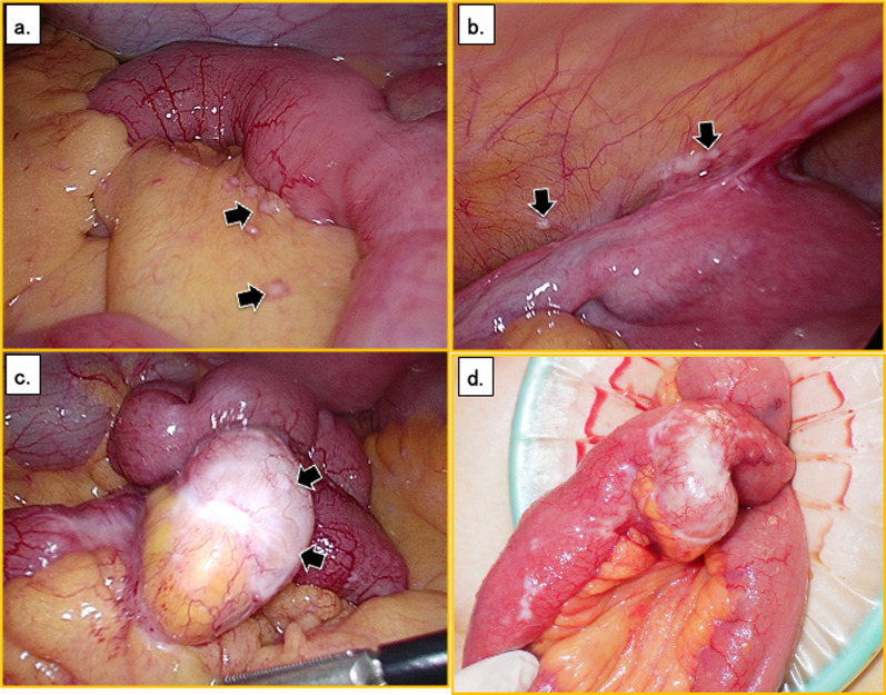
Case presentation: A woman in her sixties presented to another hospital with abdominal pain. Plain computed tomography suggested an abdominal tumor and she was referred to our hospital. Enhanced computed tomography revealed a 23-mm low-density tumor in the abdominal cavity. Surgery was performed with a tentative diagnosis of a mesenteric tumor, such as a gastrointestinal stromal tumor, schwannoma, or lymphoma. First, we inspected the peritoneal cavity with a laparoscope. This revealed numerous nodules in the small bowel mesentery, suggesting peritoneal dissemination. A 20-mm-diameter white tumor was found in the small intestine and diagnosed as a small intestinal cancer. The small intestine was partially resected laparoscopically through a small skin incision. The patient's postoperative course was uneventful, and she was discharged on postoperative day 9. Pathological examination revealed well-differentiated adenocarcinoma in the small intestine. The tumor had developed from a sac-like portion protruding toward the serosal side and had a glandular structure lined with flattened atypical cells. Neither pancreatic acinar cells nor islets of Langerhans were evident, suggesting a Heinrich type 3 ectopic pancreas. The final diagnosis was an adenocarcinoma originating from an ectopic pancreas in Meckel's diverticulum. After a smooth recovery, the patient commenced chemotherapy for pancreatic cancer.

Conclusions: We present a very rare case of ectopic pancreatic carcinoma in Meckel's diverticulum.

Keywords: Chemotherapy; Ectopic pancreatic adenocarcinoma; Meckel’s diverticulum; Rare cancer; Surgery.


Conflict of interest statement

The authors declare that they have no competing interests.

Figures

Fig. 1
Fig. 1
Enhanced computed tomography image showing a 23-mm low-density tumor in the abdominal cavity (white arrows)
Fig. 2
Fig. 2
Intraoperative findings. a There are numerous nodules in the small intestinal mesentery (black arrows). b There are also disseminated nodules in the pouch of Douglas (black arrows). c There is a 2-cm white tumor in the small intestine (black arrows). d The tumor was located approximately 130 cm on the oral side, toward the ileocecal valve
Fig. 3
Fig. 3
Histological findings with HE staining. a Photomicrograph showing a sac-like structure within the muscularis propria. The structure is a true diverticulum protruding from the small intestine (dashed yellow line). The cancerous area is shown as a Square b, and the cancer and adjacent tissue as Square c. b Photomicrograph showing atypical columnar epithelium that has grown in a tubular manner in the mucous membrane of the diverticulum. c Photomicrograph showing an enlarged space covered by flattened atypical cells in the tissue of Meckel’s diverticulum and in contact with the cancer. This tissue is suggestive of ectopic pancreas. There is no gastric mucosa, intestinal mucosa, acinar cell, or islets of Langerhans. HE, hematoxylin and eosin
Fig. 4
Fig. 4
Immunohistochemical findings for the resected cancer tissue. Photomicrographs showing a CK7 positivity; b CK20 negativity; c CDX2 negativity; and d CA19-9 positivity. CK cytokeratin, CDX2 CDX2 gene, CA19-9 carbohydrate antigen 19-9

References

    1. Leijonmarck CE, Bonman-Sandelin K, Frisell J, et al. Meckel’s diverticulum in the adult. Br J Surg. 1986;73(2):146–149. doi: 10.1002/bjs.1800730225. - DOI - PubMed
    1. Yamaguchi K, Maeda S, Kitamura K. Adenocarcinoma in Meckel’s diverticulum: case report and literature review. Aust N Z J Surg. 1989;59(10):811–813. doi: 10.1111/j.1445-2197.1989.tb07014.x. - DOI - PubMed
    1. Koh HC, Page B, Black C, et al. Ectopic pancreatic-type malignancy presenting in a Meckel’s diverticulum: a case report and review of the literature. World J Surg Oncol. 2009;7:54. doi: 10.1186/1477-7819-7-54. - DOI - PMC - PubMed
    1. Park JJ, Wolff BG, Tollefson MK, et al. Meckel diverticulum: the Mayo Clinic experience with 1476 patients (1950–2002) Ann Surg. 2005;241(3):529–533. doi: 10.1097/01.sla.0000154270.14308.5f. - DOI - PMC - PubMed
    1. Kabir SA, Raza SA, Kabir SI. Malignant neoplasms of Meckel’s diverticulum; an evidence based review. Ann Med Surg (Lond) 2019;43:75–81. doi: 10.1016/j.amsu.2019.05.017. - DOI - PMC - PubMed

LinkOut - more resources