. 2024 Jun;46(3):3481-3501.
doi: 10.1007/s11357-024-01090-7. Epub 2024 Feb 23.

Vascular smooth muscle cell-specific Igf1r deficiency exacerbates the development of hypertension-induced cerebral microhemorrhages and gait defects

Affiliations

Vascular smooth muscle cell-specific Igf1r deficiency exacerbates the development of hypertension-induced cerebral microhemorrhages and gait defects

Lauren R Miller et al. Geroscience. 2024 Jun.

Abstract

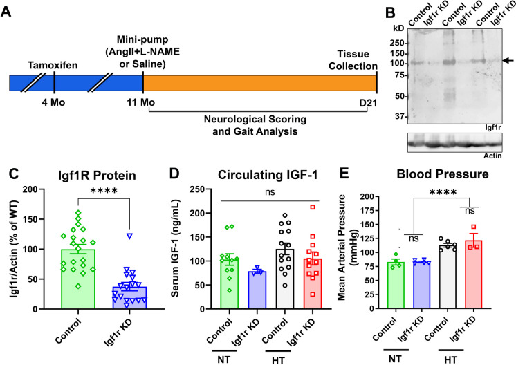
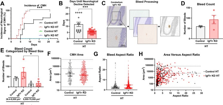
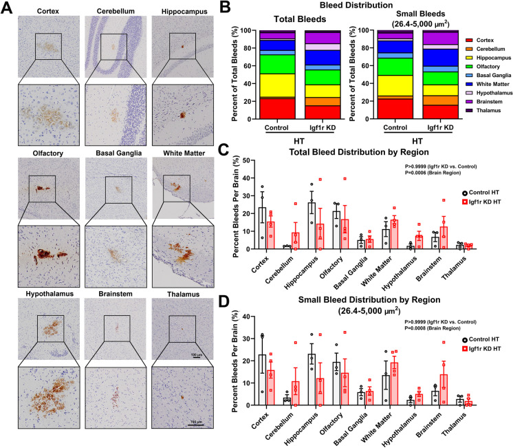
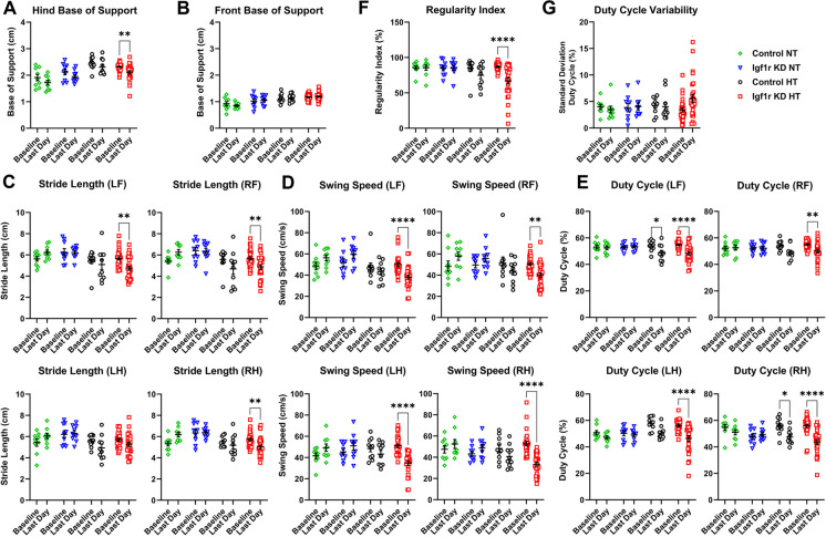
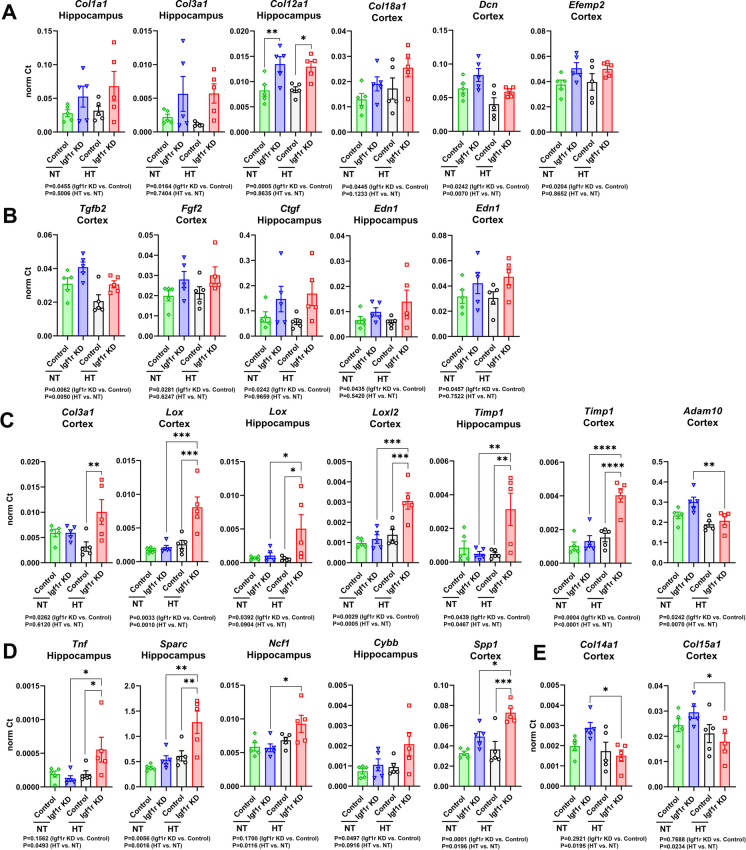
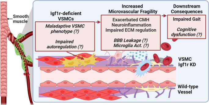
Cerebrovascular fragility and cerebral microhemorrhages (CMH) contribute to age-related cognitive impairment, mobility defects, and vascular cognitive impairment and dementia, impairing healthspan and reducing quality of life in the elderly. Insulin-like growth factor 1 (IGF-1) is a key vasoprotective growth factor that is reduced during aging. Circulating IGF-1 deficiency leads to the development of CMH and other signs of cerebrovascular dysfunction. Here our goal was to understand the contribution of IGF-1 signaling on vascular smooth muscle cells (VSMCs) to the development of CMH and associated gait defects. We used an inducible VSMC-specific promoter and an IGF-1 receptor (Igf1r) floxed mouse line (Myh11-CreERT2 Igf1rf/f) to knockdown Igf1r. Angiotensin II in combination with L-NAME-induced hypertension was used to elicit CMH. We observed that VSMC-specific Igf1r knockdown mice had accelerated development of CMH, and subsequent associated gait irregularities. These phenotypes were accompanied by upregulation of a cluster of pro-inflammatory genes associated with VSMC maladaptation. Collectively our findings support an essential role for VSMCs as a target for the vasoprotective effects of IGF-1, and suggest that VSMC dysfunction in aging may contribute to the development of CMH.

Keywords: Aging brain; Cerebral microhemorrhage; Cerebrovascular aging; Insulin-like growth factor 1; Vascular cognitive impairment; Vascular smooth muscle cells.


Conflict of interest statement

Zoltan Ungvari is the Editor-in-Chief of Geroscience. Shannon Conley, Stefano Tarantini, Adam Nyul-Toth, and Andriy Yabluchanskiy are Associate Editors of Geroscience.

Figures

Fig. 1
Fig. 1
VSMC-specific Igf1r KD model: A. Experimental paradigm for the Igf1r knockdown mouse model. B and C. Igf1r knockdown was confirmed by western blot of aortic protein extracts collected at 1 year of age in control (n = 20) and Igf1r KD (n = 17) normotensive (NT) mice. Igf1r is present ~ 100 kDa (arrow). Densitometry signal was normalized to β-actin levels. Igf1r KD animals had lower levels of Igf1r protein than their control counterparts (****P < 0.0001 by Mann–Whitney t-test). D. Serum IGF-1 levels were evaluated by ELISA (ns: not significant by two-way ANOVA with Tukey’s post hoc test) in control (n = 11) and Igf1r KD (n = 3), normotensive and control (n = 14), and Igf1r KD (n = 12) hypertensive (HT) mice. E. Mean arterial blood pressure was measured immediately prior to euthanasia (n = 4, control normotensive; n = 5, Igf1r KD normotensive; n = 6, control HT; n = 3, Igf1r KD HT). Combined treatment with AngII and L-NAME successfully increased mean arterial blood pressure in both control and Igf1r KD mice (****P < 0.0001, two-way ANOVA, with Tukey’s post hoc comparison). All graphs show mean ± SEM
Fig. 2
Fig. 2
VSMC-specific Igf1r KD accelerates development of CMH: 10–12-month-old Igf1r KD and control mice were implanted with mini-pumps containing saline (NT: normotensive) or angiotensin II (HT: hypertensive) (pump implantation was day 0) and given drinking water supplemented with L-NAME. A. Incidence curves plotting incidence of neurological signs of CMH. Igf1r KD HT mice experienced an earlier onset of neurological signs than control HT mice (incidence curves were compared using Kaplan–Meier analysis, ***P < 0.001, n = 15 control HT, n = 29 Igf1r KD HT, n = 9 control NT, n = 12 Igf1r KD NT, sample sizes the same for B). B. Igf1r KD mice developed signs of CMH significantly sooner than controls (***P < 0.001 by Mann–Whitney test). C. Serially sectioned brains from HT Igf1r KD and control animals were DAB stained (brown) and counterstained for nuclei (blue). Shown are images from the Fiji script used to analyze bleeds and collect metrics. D. Plotted is total number of bleeds per brain (Igf1r KD n = 4 animals, control n = 3) E. Bleed number was subgrouped into small bleeds (26.4–5,000 µm2) and large bleeds (5,001–70,000 µm2). Between-group differences were analyzed using two-way ANOVA followed by Tukey’s post hoc comparison, *P < 0.05. F. Plotted is bleed area (µm2) for individual bleeds in HT Igf1r KD and control animals (displayed as median ± 95% confidence interval since data were not normally distributed). No significant difference was observed between groups (P = 0.8402 by Mann–Whitney test). G. Aspect ratio was analyzed for both HT Igf1r KD and control animals (plotted as median ± 95% confidence interval). No significant difference was observed between groups (P = 0.3825 by Mann–Whitney test). H. Plotted are individual bleed areas as a function of aspect ratio. Except where otherwise noted, graphs plot mean ± SEM
Fig. 3
Fig. 3
VSMC-specific Igf1r alters bleed burden and distribution: bleeds from hypertensive Igf1r KD (n = 4) and control animals (n = 3) were mapped to nine brain regions (cortex, cerebellum, hippocampus, olfactory bulb, basal ganglia, white matter, hypothalamus, brainstem, and thalamus) to assess changes in bleed distribution across groups. A. Example images of bleeds across the nine brain regions from Igf1r KD animals. Scale bars, 100 µm. Boxed areas from top images are blown up above. Brown DAB staining marks CMH. BD. Overall bleed distribution by group is plotted for total bleeds and small bleeds. Plotted are the percentage of total bleeds (C) and small bleeds (D) bleeds found in each region for each brain. Between group differences were analyzed by two-way ANOVA (ANOVA P-values were > 0.9999 for Igf1r KD vs. control, ***P < 0.001 for brain region, no pairwise comparisons were significant in post hoc tests)
Fig. 4
Fig. 4
VSMC-specific Igf1r KD leads to gait defects in the context of CMH: prior to implantation of 10–12-month-old Igf1r KD and control mice with mini-pumps containing saline (NT: normotensive) or angiotensin II (HT: hypertensive), animals underwent baseline gait testing. Starting at day 3 post-pump implantation, animals underwent daily gait testing until removal from the study (n = 9, control normotensive; n = 9, Igf1r KD normotensive; n = 10, control hypertensive; n = 27, Igf1r KD HT). Plotted are comparisons between baseline measurements and last day (assessed by repeated measures two-way ANOVA followed by Tukey’s post hoc tests comparing baseline to last day). *P < 0.05, **P < 0.01, ****P < 0.0001. Plotted are various gait metrics, hind, and front base of support (A and B), stride length (C), swing speed (D), duty cycle (E), regularity index (F), and duty cycle variability (G) (mean ± SEM)
Fig. 5
Fig. 5
VSMC-specific Igf1r KD leads to alterations in the transcription of CMH-associated genes: at day 4 after mini-pump implantation, brains from all four groups were harvested and dissected. cDNA from cortical and hippocampal tissues (n = 5 for all groups) underwent qPCR for a panel of 96 CMH-associated genes. CT values were normalized (refer to methods). A and B. Plotted are genes upregulated in Igf1r KD animals independent of hypertension. C and D. Plotted are genes upregulated in Igf1r HT animals. E Plotted are genes downregulated in Igf1r HT animals. Graphs show means ± SEM; between-group differences were analyzed by two-way ANOVA with Tukey’s post hoc tests. P-values for two-way ANOVAs are reported underneath each graph, where post hoc tests were significant; they are indicated on the graphs with brackets, *P < 0.05, **P < 0.01, ****P < 0.0001 for post hoc tests
Fig. 6
Fig. 6
Overall proposed paradigm. Show is a depiction of our overall hypothesis. We hypothesize that Igf1r deficiency in VSMCs may lead to maladaptive VSMC phenotypic switching and impaired myogenic autoregulation. As a result, we predict increased microvascular vessel fragility, resultin CMH, and other pathologies, culminating in impaired gait and cognition. Items in regular type were evaluated in this study; italicized items with (?) remain to be evaluated in future

Similar articles

Cited by

References

    1. Gorelick PB, Counts SE, Nyenhuis D. Vascular cognitive impairment and dementia. Biochim Biophys Acta. 2016;1862(5):860–868. doi: 10.1016/j.bbadis.2015.12.015. - DOI - PMC - PubMed
    1. Gorelick PB, Scuteri A, Black SE, Decarli C, Greenberg SM, Iadecola C, et al. Vascular contributions to cognitive impairment and dementia: a statement for healthcare professionals from the American Heart Association/American Stroke Association. Stroke. 2011;42(9):2672–2713. doi: 10.1161/STR.0b013e3182299496. - DOI - PMC - PubMed
    1. Iadecola C, Duering M, Hachinski V, Joutel A, Pendlebury ST, Schneider JA, et al. Vascular cognitive impairment and dementia: JACC scientific expert panel. J Am Coll Cardiol. 2019;73(25):3326–3344. doi: 10.1016/j.jacc.2019.04.034. - DOI - PMC - PubMed
    1. Stokes AC, Weiss J, Lundberg DJ, Xie W, Kim JK, Preston SH, et al. Estimates of the association of dementia with US mortality levels using linked survey and mortality records. JAMA Neurol. 2020;77(12):1543–1550. doi: 10.1001/jamaneurol.2020.2831. - DOI - PMC - PubMed
    1. Fisher M, French S, Ji P, Kim RC. Cerebral microbleeds in the elderly: a pathological analysis. Stroke. 2010;41(12):2782–2785. doi: 10.1161/strokeaha.110.593657. - DOI - PMC - PubMed

Publication types

Substances