

Phenotypically concordant distribution of pick bodies in aphasic versus behavioral dementias
- PMID: 38389095
- PMCID: PMC10885488
- DOI: 10.1186/s40478-024-01738-7
Phenotypically concordant distribution of pick bodies in aphasic versus behavioral dementias
Abstract
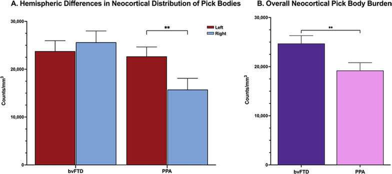
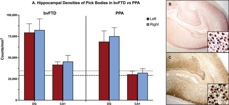
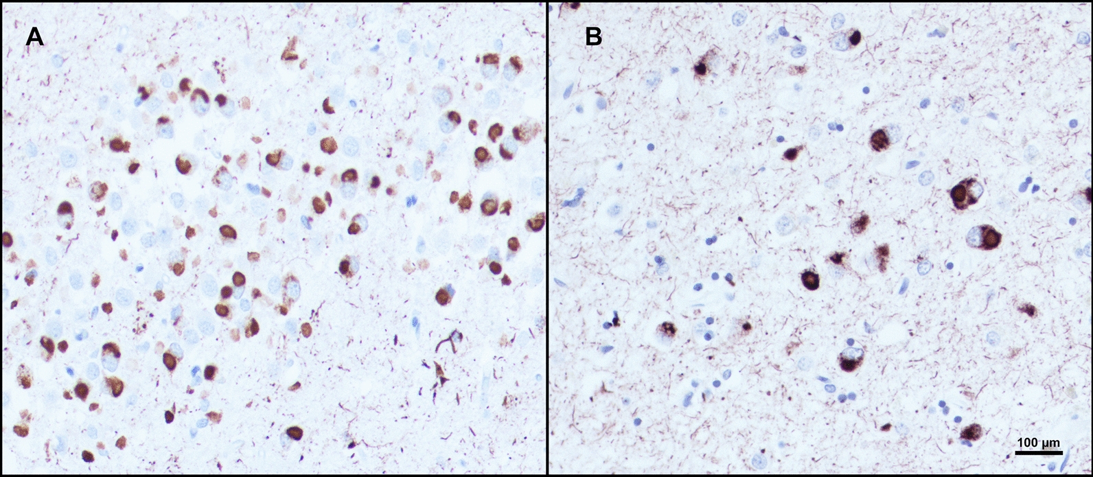
Pick's disease (PiD) is a subtype of the tauopathy form of frontotemporal lobar degeneration (FTLD-tau) characterized by intraneuronal 3R-tau inclusions. PiD can underly various dementia syndromes, including primary progressive aphasia (PPA), characterized by an isolated and progressive impairment of language and left-predominant atrophy, and behavioral variant frontotemporal dementia (bvFTD), characterized by progressive dysfunction in personality and bilateral frontotemporal atrophy. In this study, we investigated the neocortical and hippocampal distributions of Pick bodies in bvFTD and PPA to establish clinicopathologic concordance between PiD and the salience of the aphasic versus behavioral phenotype. Eighteen right-handed cases with PiD as the primary pathologic diagnosis were identified from the Northwestern University Alzheimer's Disease Research Center brain bank (bvFTD, N = 9; PPA, N = 9). Paraffin-embedded sections were stained immunohistochemically with AT8 to visualize Pick bodies, and unbiased stereological analysis was performed in up to six regions bilaterally [middle frontal gyrus (MFG), superior temporal gyrus (STG), inferior parietal lobule (IPL), anterior temporal lobe (ATL), dentate gyrus (DG) and CA1 of the hippocampus], and unilateral occipital cortex (OCC). In bvFTD, peak neocortical densities of Pick bodies were in the MFG, while the ATL was the most affected in PPA. Both the IPL and STG had greater leftward pathology in PPA, with the latter reaching significance (p < 0.01). In bvFTD, Pick body densities were significantly right-asymmetric in the STG (p < 0.05). Hippocampal burden was not clinicopathologically concordant, as both bvFTD and PPA cases demonstrated significant hippocampal pathology compared to neocortical densities (p < 0.0001). Inclusion-to-neuron analyses in a subset of PPA cases confirmed that neurons in the DG are disproportionately burdened with inclusions compared to neocortical areas. Overall, stereological quantitation suggests that the distribution of neocortical Pick body pathology is concordant with salient clinical features unique to PPA vs. bvFTD while raising intriguing questions about the selective vulnerability of the hippocampus to 3R-tauopathies.
Keywords: Behavioral variant frontotemporal dementia; Frontotemporal lobar degeneration; Pick’s disease; Primary progressive aphasia; Stereology; Tau.
© 2024. The Author(s).
Conflict of interest statement
The authors declare that they have no competing interests.
Figures


Similar articles
-
Atrophy network mapping of clinical subtypes and main symptoms in frontotemporal dementia.Brain. 2024 Sep 3;147(9):3048-3058. doi: 10.1093/brain/awae067. Brain. 2024. PMID: 38426222 Free PMC article.
-
Differential vulnerability of the dentate gyrus to tauopathies in dementias.Acta Neuropathol Commun. 2023 Jan 3;11(1):1. doi: 10.1186/s40478-022-01485-7. Acta Neuropathol Commun. 2023. PMID: 36597124 Free PMC article.
-
Itching Frequency and Neuroanatomic Correlates in Frontotemporal Lobar Degeneration.JAMA Neurol. 2024 Sep 1;81(9):977-984. doi: 10.1001/jamaneurol.2024.2213. JAMA Neurol. 2024. PMID: 39037825 Free PMC article.
-
Non-pharmacological interventions for improving language and communication in people with primary progressive aphasia.Cochrane Database Syst Rev. 2024 May 29;5(5):CD015067. doi: 10.1002/14651858.CD015067.pub2. Cochrane Database Syst Rev. 2024. PMID: 38808659 Free PMC article.
-
Regional cerebral blood flow single photon emission computed tomography for detection of Frontotemporal dementia in people with suspected dementia.Cochrane Database Syst Rev. 2015 Jun 23;2015(6):CD010896. doi: 10.1002/14651858.CD010896.pub2. Cochrane Database Syst Rev. 2015. PMID: 26102272 Free PMC article.
References
-
- Cairns NJ, Bigio EH, Mackenzie IR, Neumann M, Lee VM, Hatanpaa KJ, White CL, 3rd, Schneider JA, Grinberg LT, Halliday G, et al. Neuropathologic diagnostic and nosologic criteria for frontotemporal lobar degeneration: consensus of the Consortium for Frontotemporal Lobar Degeneration. Acta Neuropathol. 2007;114:5–22. doi: 10.1007/s00401-007-0237-2. - DOI - PMC - PubMed
Publication types
MeSH terms
Grants and funding
- K08 AG065463/AG/NIA NIH HHS/United States
- R01 DC008552/DC/NIDCD NIH HHS/United States
- P30AG013854/AG/NIA NIH HHS/United States
- R01 AG077444/AG/NIA NIH HHS/United States
- U01 AG016976/AG/NIA NIH HHS/United States
- T32 AG020506/AG/NIA NIH HHS/United States
- P30 AG072977/AG/NIA NIH HHS/United States
- R01 NS085770/NS/NINDS NIH HHS/United States
- R01 NS075075/NS/NINDS NIH HHS/United States
- R01 AG062566/AG/NIA NIH HHS/United States
- P30 AG013854/AG/NIA NIH HHS/United States
- R01 AG056258/AG/NIA NIH HHS/United States
- F31 AG076318/AG/NIA NIH HHS/United States
- T32 NS047987/NS/NINDS NIH HHS/United States
- P30 AG066546/AG/NIA NIH HHS/United States
- R56 AG075600/AG/NIA NIH HHS/United States
LinkOut - more resources
Full Text Sources
Medical
Miscellaneous
