. 2024 Feb 22;22(1):144.
doi: 10.1186/s12964-024-01523-y.

Hypoxic glioblastoma-cell-derived extracellular vesicles impair cGAS-STING activity in macrophages

Affiliations

Hypoxic glioblastoma-cell-derived extracellular vesicles impair cGAS-STING activity in macrophages

Stoyan Tankov et al. Cell Commun Signal. .

Abstract

Background: Solid tumors such as glioblastoma (GBM) exhibit hypoxic zones that are associated with poor prognosis and immunosuppression through multiple cell intrinsic mechanisms. However, release of extracellular vesicles (EVs) has the potential to transmit molecular cargos between cells. If hypoxic cancer cells use EVs to suppress functions of macrophages under adequate oxygenation, this could be an important underlying mechanism contributing to the immunosuppressive and immunologically cold tumor microenvironment of tumors such as GBM.

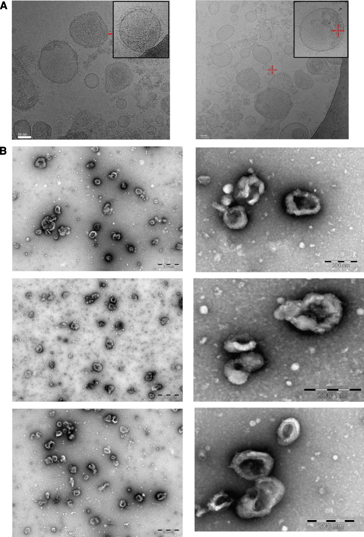
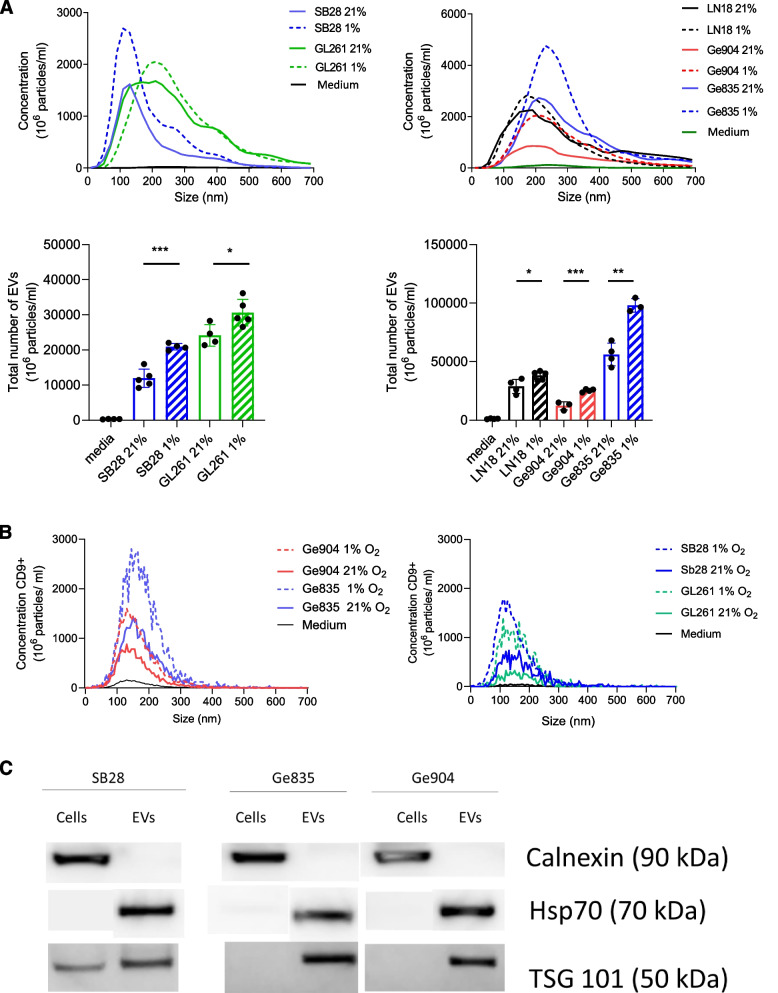
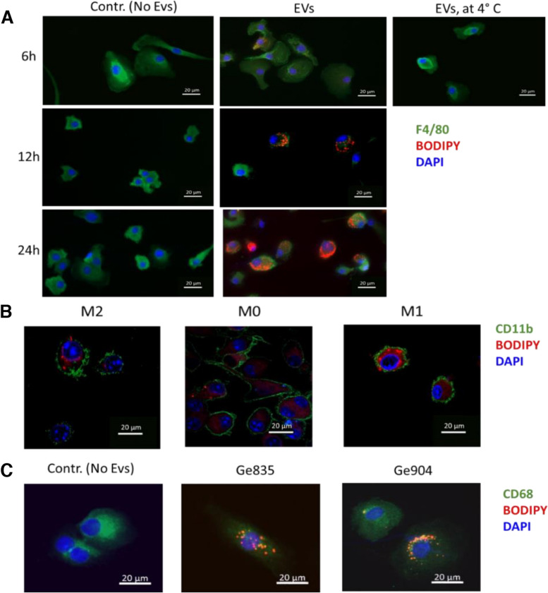
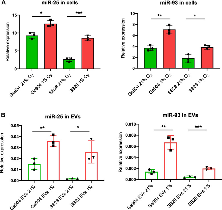
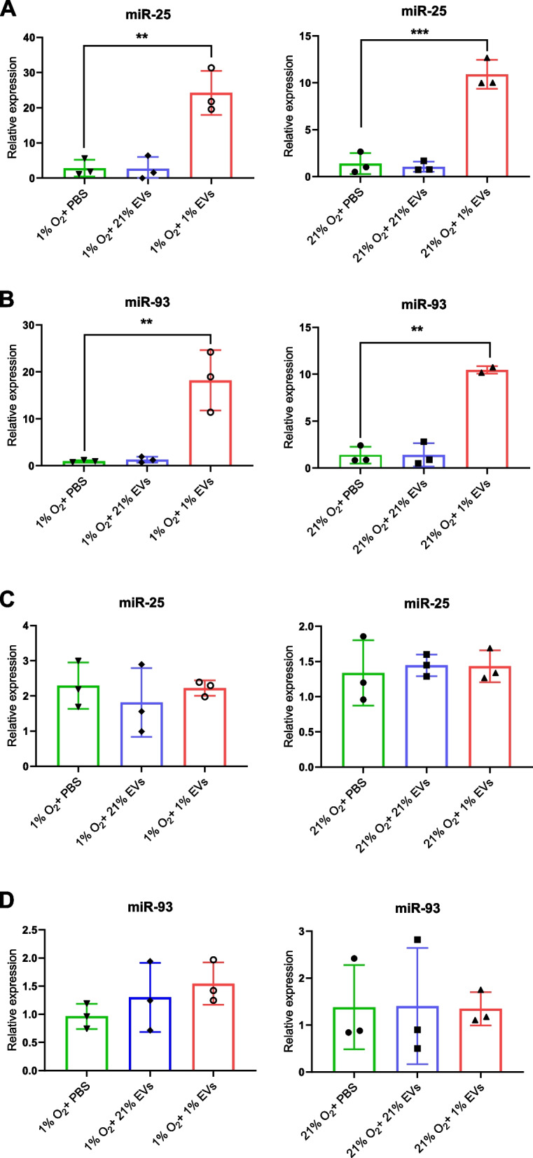
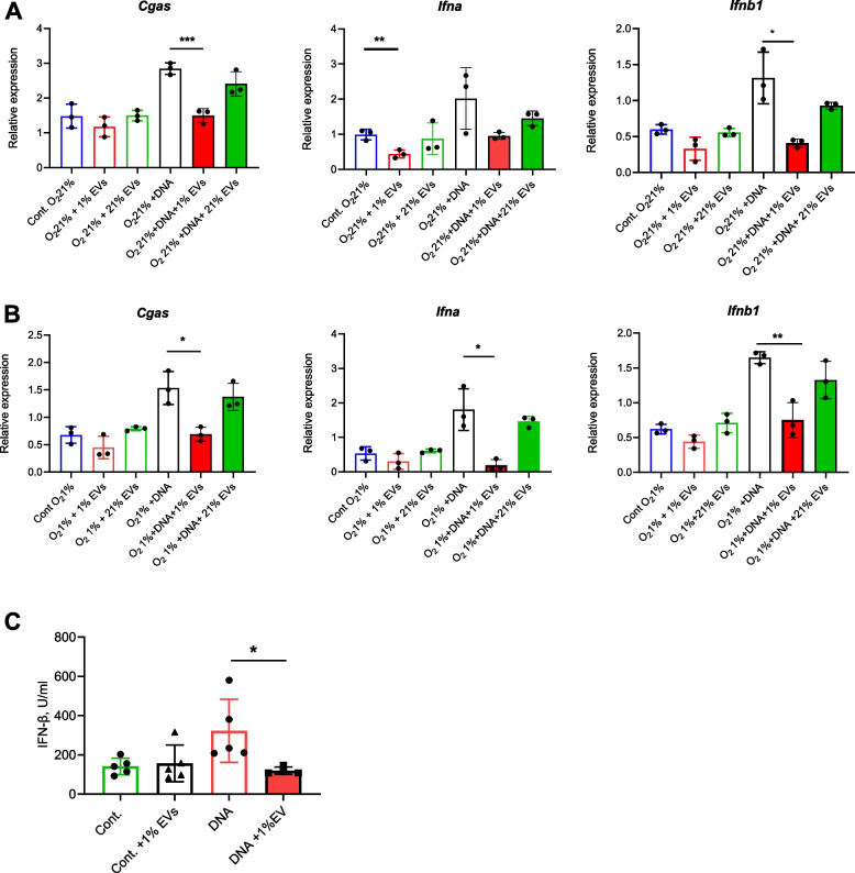
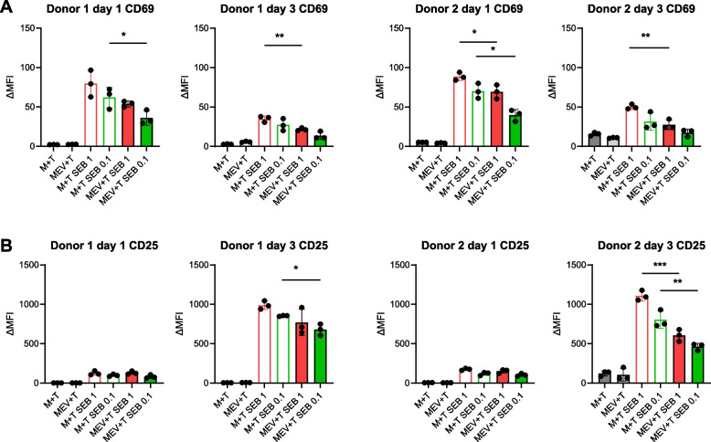
Methods: EVs were isolated by differential ultracentrifugation from GBM cell culture supernatant. EVs were thoroughly characterized by transmission and cryo-electron microscopy, nanoparticle tracking analysis (NTA), and EV marker expression by Western blot and fluorescent NTA. EV uptake by macrophage cells was observed using confocal microscopy. The transfer of miR-25/93 as an EV cargo to macrophages was confirmed by miRNA real-time qPCR. The impact of miR-25/93 on the polarization of recipient macrophages was shown by transcriptional analysis, cytokine secretion and functional assays using co-cultured T cells.

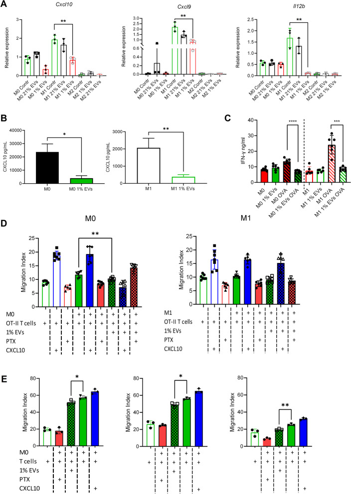
Results: We show that indirect effects of hypoxia can have immunosuppressive consequences through an EV and microRNA dependent mechanism active in both murine and human tumor and immune cells. Hypoxia enhanced EV release from GBM cells and upregulated expression of miR-25/93 both in cells and in EV cargos. Hypoxic GBM-derived EVs were taken up by macrophages and the miR-25/93 cargo was transferred, leading to impaired cGAS-STING pathway activation revealed by reduced type I IFN expression and secretion by macrophages. The EV-treated macrophages downregulated expression of M1 polarization-associated genes Cxcl9, Cxcl10 and Il12b, and had reduced capacity to attract activated T cells and to reactivate them to release IFN-γ, key components of an efficacious anti-tumor immune response.

Conclusions: Our findings suggest a mechanism by which immunosuppressive consequences of hypoxia mediated via miRNA-25/93 can be exported from hypoxic GBM cells to normoxic macrophages via EVs, thereby contributing to more widespread T-cell mediated immunosuppression in the tumor microenvironment.

Keywords: Extracellular vesicles; Glioblastoma; Hypoxia/ miRNA; cGAS-STING pathway.


Conflict of interest statement

The authors declare no competing interests.

Figures

Fig. 1
Fig. 1
Electron microscopy characterization of GBM-derived EVs from normoxic or hypoxic culture. A Electron cryo-microscopy (cryo-EM) imaging of EVs secreted by murine (SB28, left) and human (Ge904, right) GBM cell lines. Pictures are representative of at least 3 images. B Transmission Electron Microscopy (TEM) imaging of EVs secreted by murine (SB28, top row) and human (Ge904, middle row and Ge835 lower row) GBM cell lines. Pictures are representative of at least 6 images
Fig. 2
Fig. 2
NTA characterization of GBM-derived EVs from normoxic or hypoxic culture. A NTA profiles of EVs isolated from murine SB28 and GL261 cell lines (left) and from human Ge904, Ge835 and LN18 GBM cell lines (right) cultured in hypoxic (1% O2) or normoxic (21% O2) conditions. (upper panels). Total number of EVs measured by NTA from murine (SB28 and GL261) (left) and from human (Ge904, Ge835 and LN18) GBM cell lines (right) cultured in hypoxic (1% O2) or normoxic (21% O2) conditions (lower panel). EV depleted culture media was used as control. The calculated size distribution in the upper panels is depicted as a mean from three experiments and three measurements. In the lower panels, data is presented as the mean ± SD of three independent experiments and comparisons were made using an unpaired t test. *p < 0.05, **p < 0.005, ***P < 0.001. B fNTA profiles of EVs isolated from murine SB28 and GL261 cell lines (left) and from human Ge904, Ge835 and GBM cell lines (right) cultured in hypoxic (1% O2) or normoxic (21% O2) conditions. The EVs were stained with APC conjugated anti-CD9 antibody (anti-mouse or anti-human correspondingly). EV depleted culture media was used as control. The calculated size distribution is depicted as a mean from three experiments and three measurements. C Western blot analysis of cells and EVs from murine (SB28) and human (Ge904 and Ge835) cell lines
Fig. 3
Fig. 3
GBM-derived EVs are internalized by macrophages. A Kinetics of macrophage uptake of BODIPY membrane-labelled (red) EVs derived from hypoxic SB28 cells at the indicated times. EPMs were stained for F4/80 (green) and DAPI (blue). Negative control: macrophages incubated with addition of non-labelled EVs. For passive membrane uptake control, macrophages were incubated at 4 °C for 6 h with BODIPY membrane-labelled EVs. B Uptake of hypoxic SB28-derived EVs labeled with BODIPY (red) by BMDMs, M0, or after polarization to M1 and M2. BMDMs were incubated with labeled EVs for 24 h at 37 °C and stained for CD11b (green) and DAPI (blue). C Uptake of BODIPY-labeled (red) EVs from hypoxic Ge835 or Ge904 cells by human monocyte-derived macrophages (MDMs) stained for CD68 (green) and DAPI (blue). Control: MDMs without EVs. MDMs were incubated with the corresponding labeled EVs for 24 h at 37 °C before fixation and imaging. Pictures are representative of a minimum of 3 images per group
Fig. 4
Fig. 4
Hypoxia upregulates mir25/93 expression in human and mouse GBM cells and derived EVs. A Cellular expression levels of miR-25 (left) and miR-93 (right) in human (Ge904) and mouse (SB28) GBM cells measured by RT-qPCR. miR-191 was used as stably expressed housekeeping miRNA normalization control. B EVs secreted from human (Ge904) and mouse (SB28) GBM cells cultured for 24 h in hypoxic (1% O2) or normoxic (21% O2) conditions were analyzed for miR-25 (left) and miR-93(right) levels by RT-qPCR. cel-miR-39 spike-in control was added as a normalization control. Data is presented as the mean ± SD of three independent experiments and comparisons were made using an unpaired t test. *p < 0.05, **p < 0.005, ***P < 0.001
Fig. 5
Fig. 5
Hypoxic GBM cells deliver miR-25/93 to macrophages via hypoxic GBM-derived EVs. A., B. BMDMs were incubated for 24 h under hypoxia (1% O2) or normoxia (21% O2) with addition of EVs derived from hypoxic (1% EVs) or normoxic (21% EVs) SB28 cells. Total miRNA was extracted from the BMDMs and the levels of miR-25 (A) and miR-93 (B) were measured by RT-qPCR. C., D. BMDMs were incubated for 24 h under hypoxia (1% O2) or normoxia (21% O2) with addition of EVs derived from hypoxic (1% EVs) or normoxic (21% EVs) miR25/93 KO SB28 cells. Total miRNA was extracted from the BMDMs and the levels of miR-25 (C) and miR-93 (D) were measured by RT-qPCR. Values are expressed as mean ± SD of three biologic replicates, and comparisons were made using an unpaired t test. **p < 0.005, ***P < 0.001
Fig. 6
Fig. 6
Hypoxic-GBM EVs reduce cGAS and downstream type I IFN responses in macrophages. A, B mRNA expression in EPMs of genes involved in the regulation (cGAS) and production of IFN-α and IFN-β. Macrophages were co-cultured with or without hypoxic SB28-derived EVs (1% EVs) or normoxic SB28-derived EVs (21% EVs) and 5 μg/ml of total SB28 DNA (DNA) under normoxic (O2 21%) or hypoxic (O2 1%) conditions for 24 h. C Secretion levels of IFN-β protein from EPM supernatants analyzed by ELISA. Macrophages were co-cultured with or without 5 μg/ml SB28 total DNA (DNA) and EVs from hypoxic SB28 cells (1% EVs) for 24 h. Values are expressed as mean ± SD of three biologic replicates, and comparisons were made using an unpaired t test. *p < 0.05, **p < 0.005, ***P < 0.001
Fig. 7
Fig. 7
Hypoxic GBM-derived EVs disrupt T cell attraction and reactivation capacity of macrophages. A-D Mouse BMDMs were unpolarized (M0), M1 polarized (M1), or M2 polarized (M2) for 7 days in the absence (contr.) or presence of hypoxic GBM-derived EVs (1% EVs) or normoxic GBM-derived EVs (21% EVs). A mRNA expression of Cxcl9, Cxcl10 and Il12b in BMDMs. mRNA levels were measured by RT-qPCR and expression was normalized to housekeeping genes (Gapdh and Eef1a1). B Macrophages were harvested, washed, then incubated for a further 24 h. Supernatants were collected and CXCL10 secretion was measured by a flow cytometry bead-based assay. C Macrophages were harvested, washed, pulsed with OVA peptide then added to OT-II CD4+ cells. IFN-γ secretion was measured after 24 h by ELISA. D (Left panel) Macrophages were harvested, washed, then added to the lower chambers of Transwell plates for 3 h. CD4+ OT-II T cells were added to the upper chamber and migration measured after 6 h. Negative migration control: CD4+ T cells were pretreated with pertussis toxin (PTX). Positive migration control: CXCL10 was added to the BMDMs in the lower chamber (CXCL10). E (Left, center and right panel) Human MDMs (M0) from three donors were harvested, washed, then added to the lower chambers of Transwell plates for 3 h. (Left and center) CD3+ T cells from the corresponding buffy coat donor were added to the upper chamber and the migration was measured after 6 h. Negative migration control: CD3+ T cells were pretreated with pertussis toxin (PTX). Positive migration control: CXCL10 was added to the human macrophages in the lower chamber. (Right panel) Identical experiment as in left and center panels with the use of CD3+ T cells from a different donor. Values are expressed as mean ± SD of three biologic replicates, and comparisons were made using an unpaired t test. *p < 0.05, **p < 0.005, ***P < 0.001, ****P < 0.0001
Fig. 8
Fig. 8
Human macrophages treated with hypoxic GBM-derived EVs have lower capacity to induce T cell activation markers after superantigen stimulation. A., B. Human MDMs from two donors (Donor 1 and Donor 2) were cultured for 7 days in the absence (M) or presence (MEV) of hypoxic GBM-derived EVs (1% EVs). The macrophages were then washed and co-cultured with CD3+ T cells from the same donor for 3 days with or without the addition of Staphylococcal Enterotoxin B (1 μg/ml, SEB 1 or 0.1 μg/ml, SEB 0.1). On day 1 and day 3 cells were harvested and the expression of (A) CD69 and (B) CD25 on T cells (CD4+ and CD8+) was measured by flow cytometry. Values are expressed as mean ± SD of three replicates for each donor, and comparisons were made using an unpaired t test. *p < 0.05, **p < 0.005, ***P < 0.001

Similar articles

Cited by

References

    1. Louis DN, Perry A, Wesseling P, Brat DJ, Cree IA, Figarella-Branger D, Hawkins C, Ng HK, Pfister SM, Reifenberger G, et al. The 2021 WHO classification of tumors of the central nervous system: a summary. Neuro-Oncology. 2021;23:1231–1251. - PMC - PubMed
    1. Calvo Tardon M, Marinari E, Migliorini D, Bes V, Tankov S, Charrier E, McKee TA, Dutoit V, Dietrich PY, Cosset E, Walker PR. An experimentally defined hypoxia gene signature in glioblastoma and its modulation by metformin. Biology (Basel) 2020;9(9):264. - PMC - PubMed
    1. Vuillefroy de Silly R, Dietrich PY, Walker PR. Hypoxia and antitumor CD8(+) T cells: an incompatible alliance? Oncoimmunology. 2016;5:e1232236. - PMC - PubMed
    1. Maire CL, Fuh MM, Kaulich K, Fita KD, Stevic I, Heiland DH, Welsh JA, Jones JC, Gorgens A, Ricklefs T, et al. Genome-wide methylation profiling of glioblastoma cell-derived extracellular vesicle DNA allows tumor classification. Neuro-Oncology. 2021;23:1087–1099. - PMC - PubMed
    1. Cianciaruso C, Beltraminelli T, Duval F, Nassiri S, Hamelin R, Mozes A, Gallart-Ayala H, Ceada Torres G, Torchia B, Ries CH, et al. Molecular profiling and functional analysis of macrophage-derived tumor extracellular vesicles. Cell Rep. 2019;27(3062–3080):e3011. - PMC - PubMed

Publication types