

Saikosaponin D alleviates inflammatory response of osteoarthritis and mediates autophagy via elevating microRNA-199-3p to target transcription Factor-4
- PMID: 38389105
- PMCID: PMC10882832
- DOI: 10.1186/s13018-024-04607-0
Saikosaponin D alleviates inflammatory response of osteoarthritis and mediates autophagy via elevating microRNA-199-3p to target transcription Factor-4
Abstract
Objective: This study was to investigate the underlying mechanism by which Saikosaponin D (SSD) mitigates the inflammatory response associated with osteoarthritis (OA) and regulates autophagy through upregulation of microRNA (miR)-199-3p and downregulation of transcription Factor-4 (TCF4).
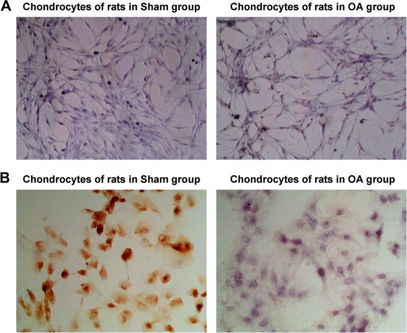
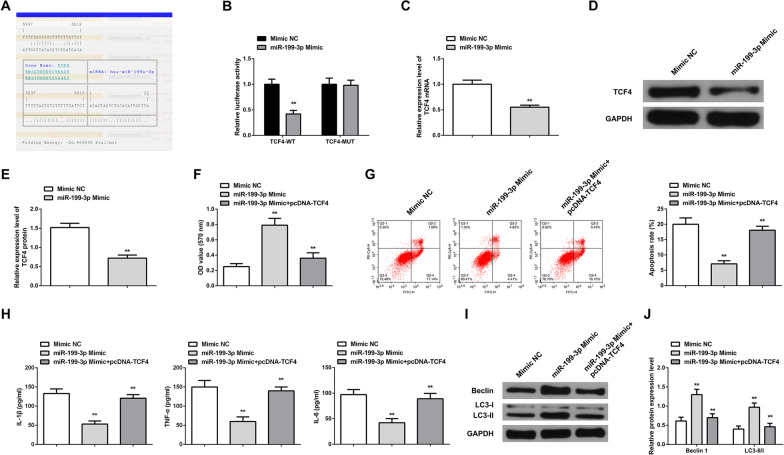
Methods: A mouse OA model was established. Mice were intragastrically administered with SSD (0, 5, 10 μmol/L) or injected with miR-199-3p antagomir into the knee. Then, pathological changes in cartilage tissues were observed. Normal chondrocytes and OA chondrocytes were isolated and identified. Chondrocytes were treated with SSD and/or transfected with oligonucleotides or plasmid vectors targeting miR-199-3p and TCF4. Cell viability, apoptosis, inflammation, and autophagy were assessed. miR-199-3p and TCF4 expressions were measured, and their targeting relationship was analyzed.
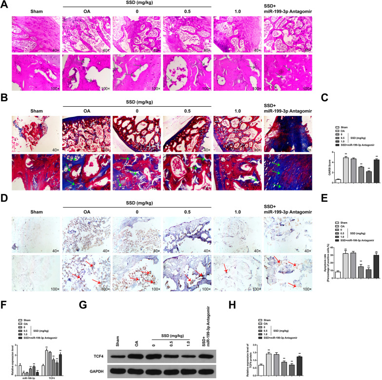
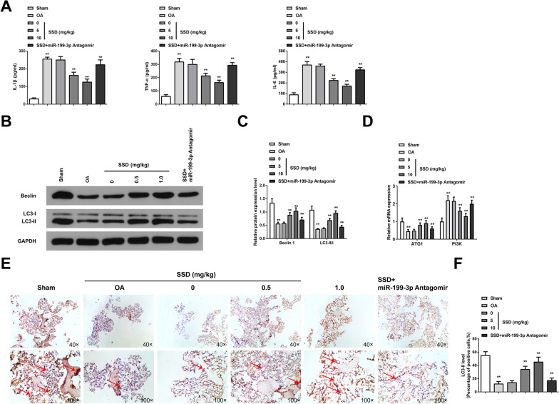
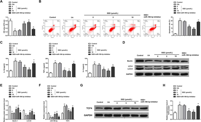
Results: In in vivo experiments, SSD ameliorated cartilage histopathological damage, decreased inflammatory factor content and promoted autophagy in OA mice. miR-199-3p expression was downregulated and TCF4 expression was upregulated in cartilage tissues of OA mice. miR-199-3p expression was upregulated and TCF4 expression was downregulated after SSD treatment. Downregulation of miR-199-3p attenuated the effect of SSD on OA mice. In in vitro experiments, SSD inhibited the inflammatory response and promoted autophagy in OA chondrocytes. Downregulation of miR-199-3p attenuated the effect of SSD on OA chondrocytes. In addition, upregulation of miR-199-3p alone inhibited inflammatory responses and promoted autophagy in OA chondrocytes. miR-199-3p targeted TCF4. Upregulation of TCF4 attenuated the effects of miR-199-3p upregulation on OA chondrocytes.
Conclusions: SSD alleviates inflammatory response and mediates autophagy in OA via elevating miR-199-3p to target TCF4.
Keywords: Autophagy; MicroRNA-42; Osteoarthritis; Saikosaponin D; Transcription Factor-4.
© 2024. The Author(s).
Conflict of interest statement
Authors declared no conflict of interest.
Figures
Similar articles
-
CircRNA circ-IQGAP1 Knockdown Alleviates Interleukin-1β-Induced Osteoarthritis Progression via Targeting miR-671-5p/TCF4.Orthop Surg. 2021 May;13(3):1036-1046. doi: 10.1111/os.12923. Epub 2021 Mar 5. Orthop Surg. 2021. PMID: 33675175 Free PMC article.
-
Knockdown of PVT1 inhibits IL-1β-induced injury in chondrocytes by regulating miR-27b-3p/TRAF3 axis.Int Immunopharmacol. 2020 Feb;79:106052. doi: 10.1016/j.intimp.2019.106052. Epub 2019 Dec 18. Int Immunopharmacol. 2020. PMID: 31863917
-
Hsa_circular RNA_0045474 Facilitates Osteoarthritis Via Modulating microRNA-485-3p and Augmenting Transcription Factor 4.Mol Biotechnol. 2024 May;66(5):1174-1187. doi: 10.1007/s12033-023-01019-z. Epub 2024 Jan 11. Mol Biotechnol. 2024. PMID: 38206529
-
Knockdown of lncRNA MFI2-AS1 inhibits lipopolysaccharide-induced osteoarthritis progression by miR-130a-3p/TCF4.Life Sci. 2020 Jan 1;240:117019. doi: 10.1016/j.lfs.2019.117019. Epub 2019 Oct 31. Life Sci. 2020. PMID: 31678554 Review.
-
MicroRNAs and Autophagy: Fine Players in the Control of Chondrocyte Homeostatic Activities in Osteoarthritis.Oxid Med Cell Longev. 2017;2017:3720128. doi: 10.1155/2017/3720128. Epub 2017 Jun 21. Oxid Med Cell Longev. 2017. PMID: 28713485 Free PMC article. Review.
Cited by
-
Research progress on the molecular mechanisms of Saikosaponin D in various diseases (Review).Int J Mol Med. 2025 Mar;55(3):37. doi: 10.3892/ijmm.2024.5478. Epub 2024 Dec 24. Int J Mol Med. 2025. PMID: 39717942 Free PMC article. Review.
-
Saikosaponins alleviate depression-like behaviors of chronic unpredictable mild stress exposed mice through ERK signaling pathway.Exp Brain Res. 2025 Feb 12;243(3):63. doi: 10.1007/s00221-025-07011-0. Exp Brain Res. 2025. PMID: 39937248
-
Epigenetic mechanisms of Nsd1-mediated histone methylation modifications in chondrocyte ferroptosis in knee osteoarthritis.Biomol Biomed. 2025 Mar 7;25(4):894-904. doi: 10.17305/bb.2024.10879. Biomol Biomed. 2025. PMID: 39217430 Free PMC article.
-
Regulation of ferroptosis in osteoarthritis and osteoarthritic chondrocytes by typical MicroRNAs in chondrocytes.Front Med (Lausanne). 2024 Nov 5;11:1478153. doi: 10.3389/fmed.2024.1478153. eCollection 2024. Front Med (Lausanne). 2024. PMID: 39564502 Free PMC article. Review.
-
Chondrocyte autophagy mechanism and therapeutic prospects in osteoarthritis.Front Cell Dev Biol. 2024 Oct 23;12:1472613. doi: 10.3389/fcell.2024.1472613. eCollection 2024. Front Cell Dev Biol. 2024. PMID: 39507422 Free PMC article. Review.
References
-
- Glyn-Jones S, Palmer AJR, Agricola R, et al. Osteoarthritis Lancet. 2015;386:376–387. - PubMed
-
- Quicke J G, Conaghan P G, Corp N et al. Osteoarthritis year in review 2021: epidemiology & therapy. Osteoarthritis Cartilage. 2021; - PubMed
-
- Bannuru RR, Osani MC, Vaysbrot EE, et al. OARSI guidelines for the non-surgical management of knee, hip, and polyarticular osteoarthritis. Osteoarthritis Cartilage. 2019;27:1578–1589. - PubMed
MeSH terms
Substances
LinkOut - more resources
Full Text Sources
Medical
