

Safety and efficacy of McCarey-Kaufman medium supplemented with colistin (polymyxin E) and amphotericin B in inhibiting the multidrug-resistant Pseudomonas aeruginosa using an ex vivo donor corneal infection model
- PMID: 38389253
- PMCID: PMC11338415
- DOI: 10.4103/IJO.IJO_2616_23
Safety and efficacy of McCarey-Kaufman medium supplemented with colistin (polymyxin E) and amphotericin B in inhibiting the multidrug-resistant Pseudomonas aeruginosa using an ex vivo donor corneal infection model
Abstract
Purpose: This study aimed to evaluate the efficacy and safety of McCarey-Kaufman (MK) medium supplemented with colistin and amphotericin B in inhibiting the growth of multidrug-resistant Pseudomonas (P.) aeruginosa , using an ex vivo experimental model with human donor corneas.
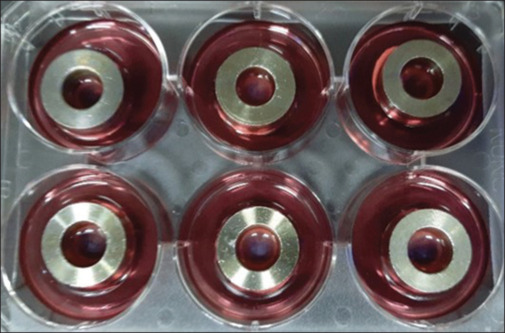
Methods: Cadaveric human corneas deemed unsuitable for corneal transplantation were obtained, and MK media were supplemented with colistin and amphotericin B. Multidrug-resistant P. aeruginosa was cultured and used to infect the human donor corneas ex vivo . Infected corneas were placed in the MK media with additional antibiotics (colistin and amphotericin B) and the standard MK media, which served as the control arm for comparison. Corneal opacity due to infiltration and quantitative analysis of colony-forming units (CFUs) were assessed. The viability of the corneal endothelium was assessed using trypan blue staining.
Results: Corneas incubated in MK media supplemented with additional antibiotics showed less corneal opacification compared with those in standard MK media at both 48- and 96-hour (hr) time points. Quantitative analysis revealed a lower bacterial load and a significant reduction in CFU in the corneas incubated in MK media with additional antibiotics compared with the control group. At 48 hrs, there was 84% ( P value = 0.024) reduction in bacterial load, and at 96 hr, a 53% ( P value = 0.016) reduction was observed in comparison with those placed in standard MK media. The trypan blue staining tests revealed that the extent of endothelial cell loss in corneas incubated in supplemented MK media was comparable to the ones in standard MK media.
Conclusion: The addition of colistin and amphotericin B to MK media demonstrated efficacy in inhibiting the growth of multidrug-resistant P. aeruginosa in an ex vivo cornea infection model. The supplemented media had no detrimental effect on the corneal endothelium. The findings suggest that supplementing the MK media with these broad-spectrum antimicrobial agents may help mitigate the risk of postoperative donor-related infection in the recipients by reducing and containing the load of microbial contamination in donor corneas.
Copyright © 2024 Copyright: © 2024 Indian Journal of Ophthalmology.
Conflict of interest statement
There are no conflicts of interest.
Figures


References
-
- Hassan SS, Wilhelmus KR. Medical review subcommittee of the Eye Bank Association of America. Eye-banking risk factors for fungal endophthalmitis compared with bacterial endophthalmitis after corneal transplantation. Am J Ophthalmol. 2005;139:685–90. - PubMed
-
- Kloess PM, Stulting RD, Waring GO, 3rd, Wilson LA. Bacterial and fungal endophthalmitis after penetrating keratoplasty. Am J Ophthalmol. 1993;115:309–16. - PubMed
-
- Taban M, Behrens A, Newcomb RL, Nobe MY, McDonnell PJ. Incidence of acute endophthalmitis following penetrating keratoplasty: A systematic review. Arch Ophthalmol. 2005;123:605–9. - PubMed
-
- Aiello LP, Javitt JC, Canner JK. National outcomes of penetrating keratoplasty. Risks of endophthalmitis and retinal detachment. Arch Ophthalmol. 1993;111:509–13. - PubMed
MeSH terms
Substances
LinkOut - more resources
Full Text Sources
Medical
