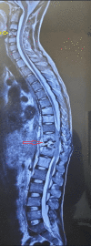
Single Sequence Whole-Spine Screening Magnetic Resonance Imaging: Diagnostic and Therapeutic Role in Multiple-Level Spinal Tuberculosis
- PMID: 38389615
- PMCID: PMC10882150
- DOI: 10.7759/cureus.52757
Single Sequence Whole-Spine Screening Magnetic Resonance Imaging: Diagnostic and Therapeutic Role in Multiple-Level Spinal Tuberculosis
Abstract
Introduction: Spinal tuberculosis (TB) is the most common form of skeletal tuberculosis. Paradiscal continuous vertebral involvement at a single level is the most prevalent pattern among all forms of spinal TB. There is a wide range of reported incidences of multiple-level non-contiguous spinal TB in the literature. We would like to discuss on the utility of single whole spine screening T2-weighted (T2W) mid-sagittal magnetic resonance imaging (MRI) film in diagnosing multiple-level spinal TB and therapeutic benefits it can provide.
Methods: We have done a retrospective review of the collected data of patients in Vardhman Mahavir Medical College and Safdarjung Hospital from August 2017 to October 2021 to find the incidence of multiple-level spinal TB and possible factors attributed to this specific disease pattern. All the patients who had been diagnosed of spinal TB either microbiologically or histopathologically or by a good clinical response to anti-tubercular treatment (ATT) and had a whole spine screening MRI film, were included. Patients of spinal TB who did not have a whole spine screening MRI were excluded from the study. Multiple-level spinal TB was diagnosed when lesions were identified in vertebral levels other than a typical paradiscal lesion, and additional lesions were separated from the primary disease by at least one normal spinal segment.
Results: Among the patients, 242 met the inclusion criteria, and 76 showed multiple-level non-contiguous spinal TB on MRI, incidence being 31.4%. The rest of the 166 patients showed typical single-segment contiguous lesions. By doing multivariate analysis to determine the independent risk factors for multiple-level spinal TB, extremes of age (<20 years and >50 years) have been found to be a significant factor with p value of 0.0001. Though drug resistance was not found to be a significant risk factor (p value 0.051), the proportion of patients having multiple-level TB was far more in the drug-resistant group (13/76).
Conclusions: Single sequence whole spine screening MRI film is an effective, economical, and time-saving tool to detect multiple-level spinal TB. Along with its diagnostic accuracy, it also provides therapeutic benefits like access to a more approachable site for biopsy.
Keywords: drug resistant spinal tuberculosis; multiple level spinal tuberculosis; spinal tuberculosis:; spine biopsy; whole spine screening mri.
Copyright © 2024, Sareen et al.
Conflict of interest statement
The authors have declared that no competing interests exist.
Figures


Similar articles
-
Role of Whole-Spine Screening Magnetic Resonance Imaging Using Short Tau Inversion Recovery or Fat-Suppressed T2 Fast Spin Echo Sequences for Detecting Noncontiguous Multiple-Level Spinal Tuberculosis.Asian Spine J. 2018 Aug;12(4):686-690. doi: 10.31616/asj.2018.12.4.686. Epub 2018 Jul 27. Asian Spine J. 2018. PMID: 30060377 Free PMC article.
-
Diagnostic accuracy of whole spine magnetic resonance imaging in spinal tuberculosis validated through tissue studies.Eur Spine J. 2019 Dec;28(12):3003-3010. doi: 10.1007/s00586-019-06031-z. Epub 2019 Jun 14. Eur Spine J. 2019. PMID: 31201566
-
Noncontiguous spinal tuberculosis: incidence and management.Eur Spine J. 2009 Aug;18(8):1096-101. doi: 10.1007/s00586-009-0966-0. Epub 2009 Apr 9. Eur Spine J. 2009. PMID: 19357878 Free PMC article.
-
Pott's Spine: Diagnostic Imaging Modalities and Technology Advancements.N Am J Med Sci. 2013 Jul;5(7):404-11. doi: 10.4103/1947-2714.115775. N Am J Med Sci. 2013. PMID: 24020048 Free PMC article. Review.
-
Skipped multifocal extensive spinal tuberculosis involving the whole spine: A case report and literature review.Medicine (Baltimore). 2018 Jan;97(3):e9692. doi: 10.1097/MD.0000000000009692. Medicine (Baltimore). 2018. PMID: 29505022 Free PMC article. Review.
References
-
- Tuberculosis elimination: what’s to stop us? Reichman LB. https://www.ingentaconnect.com/contentone/iuatld/ijtld/1997/00000001/000.... Int J Tuberc Lung Dis. 1997;1:3–11. - PubMed
-
- Tuli SM. New Delhi: Jaypee Brothers Medical Publishers. New Delhi: Jaypee Brothers Medical Publishers; 2016. Tuberculosis of the skeletal system.
LinkOut - more resources
Full Text Sources
Miscellaneous
