

Atypical Ocular Toxoplasmosis With Remote Vasculitis and Kyrieleis Plaques
- PMID: 38389616
- PMCID: PMC10882149
- DOI: 10.7759/cureus.52756
Atypical Ocular Toxoplasmosis With Remote Vasculitis and Kyrieleis Plaques
Abstract
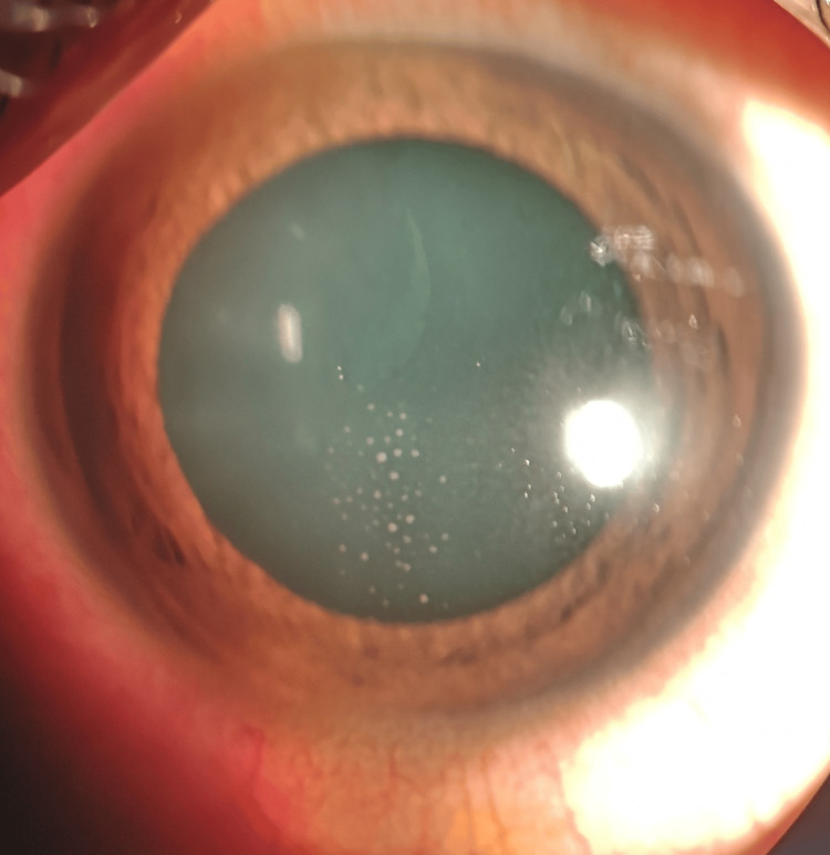
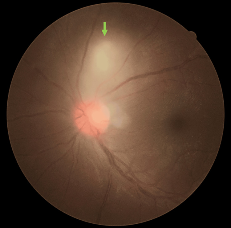
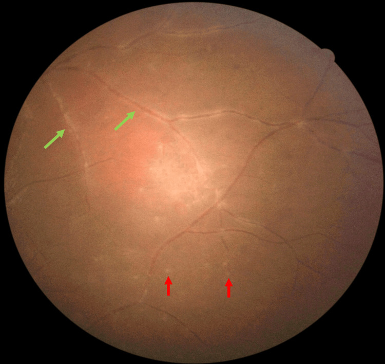
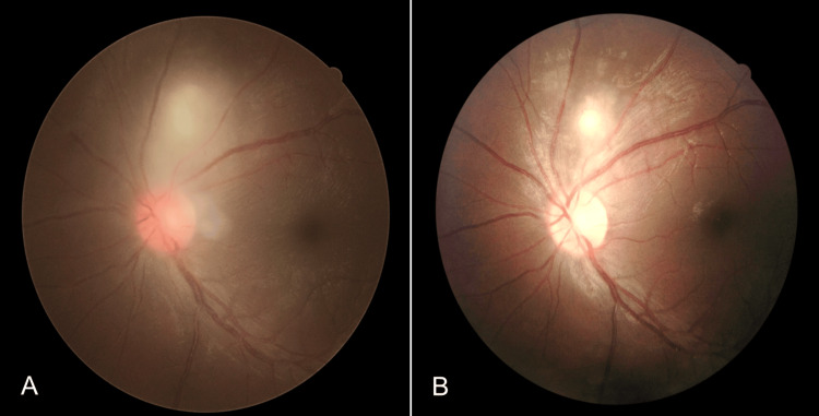
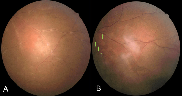
Retinal vasculitis is common in ocular toxoplasmosis (OT) and typically occurs in the same quadrant as retinochoroiditis. This is a case of atypical ocular toxoplasmosis with remote vasculitis distant from the retinochoroiditis lesion. Examination of the left fundus showed the classic posterior segment finding of "headlight in the fog" in the absence of a chorioretinal scar. Retinal vasculitis was noted in all four quadrants at the periphery far from the retinitis area. A presumptive diagnosis of acute panuveitis secondary to ocular toxoplasmosis was made despite the enzyme-linked immunosorbent assay (ELISA) for Toxoplasmosis antibody being pending. The patient was treated empirically with oral sulfamethoxazole-trimethoprim for eight weeks and received both oral and topical corticosteroids. His symptoms and ocular signs have significantly improved. This case report highlights an atypical remote localization of vasculitis with the classic appearance of retinochoroiditis and vitritis, which is highly due to toxoplasmosis. Early initiation of antibiotic therapy is recommended despite pending serology to ensure a good final visual and ocular outcome.
Keywords: atypical; atypical ocular toxoplasmosis; kyrieleis plaques; ocular toxoplasmosis; remote vasculitis.
Copyright © 2024, Teng Siew et al.
Conflict of interest statement
The authors have declared that no competing interests exist.
Figures
Similar articles
-
Unilateral vitritis in an immunocompetent individual - A rare presentation of ocular toxoplasmosis.Med J Malaysia. 2025 Jan;80(Suppl 1):17-19. Med J Malaysia. 2025. PMID: 39773937
-
Prospective randomized trial of trimethoprim/sulfamethoxazole versus pyrimethamine and sulfadiazine in the treatment of ocular toxoplasmosis.Ophthalmology. 2005 Nov;112(11):1876-82. doi: 10.1016/j.ophtha.2005.05.025. Epub 2005 Sep 19. Ophthalmology. 2005. PMID: 16171866 Clinical Trial.
-
Kyrieleis like plaques - atypical presentation of ocular Behcet's disease.Rom J Ophthalmol. 2021 Oct-Dec;65(4):383-385. doi: 10.22336/rjo.2021.75. Rom J Ophthalmol. 2021. PMID: 35087981 Free PMC article.
-
Ocular impairment of toxoplasmosis.Parassitologia. 2008 Jun;50(1-2):35-6. Parassitologia. 2008. PMID: 18693554 Review.
-
Atypical presentations of ocular toxoplasmosis.Curr Opin Ophthalmol. 2002 Dec;13(6):387-92. doi: 10.1097/00055735-200212000-00008. Curr Opin Ophthalmol. 2002. PMID: 12441842 Review.
References
-
- Ocular Toxoplasmosis. Cunningham ET Jr, Belfort R Jr, Muccioli C, Arevalo JF, Zierhut M. Ocul Immunol Inflamm. 2015;23:191–193. - PubMed
-
- Pathogenesis of ocular toxoplasmosis. Smith JR, Ashander LM, Arruda SL, et al. Prog Retin Eye Res. 2021;81:100882. - PubMed
-
- Ocular toxoplasmosis: a global reassessment. Part I: epidemiology and course of disease. Holland GN. Am J Ophthalmol. 2003;136:973–988. - PubMed
-
- Toxoplasmosis. THE LANCET. J G Montoya OL. 2004;363:1965–1973. - PubMed
Publication types
LinkOut - more resources
Full Text Sources