

The Histopathological Spectrum of Scrotal Lesions in a Tertiary Care Hospital: A Cross-Sectional Study
- PMID: 38389620
- PMCID: PMC10882216
- DOI: 10.7759/cureus.52767
The Histopathological Spectrum of Scrotal Lesions in a Tertiary Care Hospital: A Cross-Sectional Study
Abstract
Background: The incidence and clinical presentation of testicular and paratesticular lesions are variable. A preoperative diagnosis is often difficult with only a clinical examination. The diagnosis of testicular lesions is mainly based on histological investigation, despite advances in imaging and tumor marker testing. This study aimed to document the histopathological spectrum of scrotal lesions, including testicular and paratesticular lesions.
Aim: The study aimed to research the histopathological spectrum of scrotal lesions.
Settings and design: This was a cross-sectional study conducted at NKP Salve Institute of Medical Sciences & Research Centre and Lata Mangeshkar Hospital, a tertiary care hospital in Nagpur, India.
Materials and methods: Following the institutional ethics committee's approval, a two-year cross-sectional study was carried out in the tertiary care hospital. Seventy operated scrotal specimens sent for histopathological examination were included in the study. The clinical details and investigations of the patients, as well as the gross and histopathological findings of all the specimens, were studied carefully.
Statistical analysis: The clinical details and gross and histopathological findings were noted in a proforma, entered in a Microsoft Excel sheet (Microsoft Corp., Redmond, WA), and verified. The data were presented in a tabular form using tablets, pie charts, and bar diagrams. The collected data were analyzed and presented in percentages and frequencies.
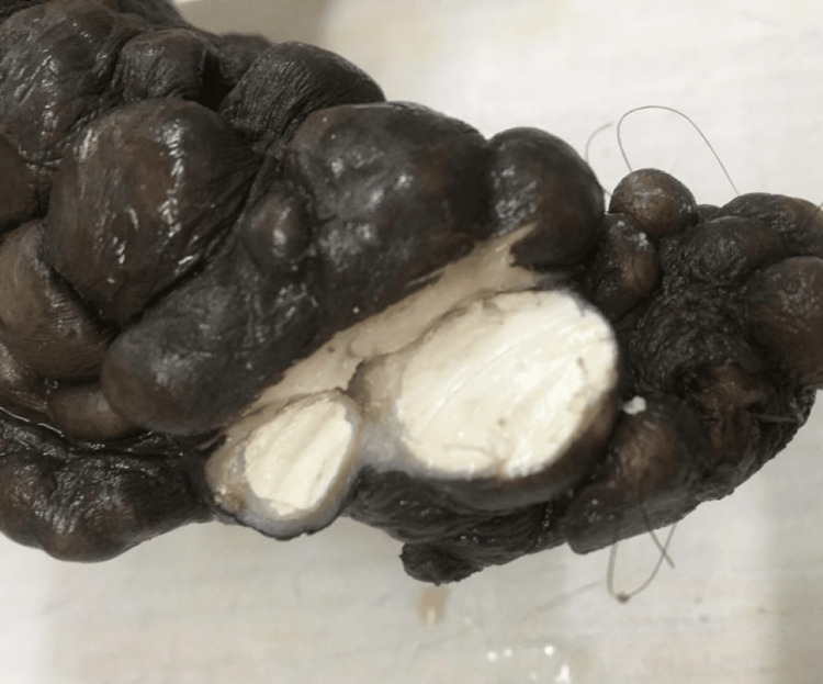
Results: The present study evaluated the histopathological spectrum of scrotal lesions in 70 operated scrotal masses. The mean age of the participants in the study was 46.55 ± 18.69 years, with the youngest patient at four years and the oldest being 88 years of age. Sixty-six (80%) of the 70 cases were of non-neoplastic lesions, while 14 (20%) were of neoplastic lesions. Testicular atrophy (16 cases) was the most common non-neoplastic lesion. The most frequent neoplastic lesion in the present study was a seminoma (seven cases).
Conclusion: This study strongly recommends routine histopathological examination of all scrotal specimens for the detection of various testicular and paratesticular lesions, as well as neoplasms. Histopathology not only provides a tissue diagnosis in scrotal disorders, but it also adds to understanding etiopathogenesis and can aid in the development of future treatment options.
Keywords: histopathological spectrum; para-testicular lesions; scrotal lesions; seminoma; testicular tumours.
Copyright © 2024, Desai et al.
Conflict of interest statement
The authors have declared that no competing interests exist.
Figures


Similar articles
-
Histopathological Trends of Testicular Neoplasm: An Experience over a Decade in a Tertiary Care Centre in the Malwa Belt of Central India.J Clin Diagn Res. 2016 Jun;10(6):EC16-8. doi: 10.7860/JCDR/2016/18238.8025. Epub 2016 Jun 1. J Clin Diagn Res. 2016. PMID: 27504294 Free PMC article.
-
Histopathological Spectrum of Tumor and Tumor-like Lesions of the Paratestis in a Tertiary Care Hospital.Oman Med J. 2015 Nov;30(6):461-8. doi: 10.5001/omj.2015.90. Oman Med J. 2015. PMID: 26674546 Free PMC article.
-
Testicular and paratesticular pathology in children: a 12-year histopathological review.World J Surg. 2010 May;34(5):969-74. doi: 10.1007/s00268-010-0459-7. World J Surg. 2010. PMID: 20151127
-
Unusual scrotal pathology: an overview.Nat Rev Urol. 2009 Sep;6(9):491-500. doi: 10.1038/nrurol.2009.149. Epub 2009 Aug 11. Nat Rev Urol. 2009. PMID: 19668251 Review.
-
Spectrum of Extratesticular and Testicular Pathologic Conditions at Scrotal MR Imaging.Radiographics. 2018 May-Jun;38(3):806-830. doi: 10.1148/rg.2018170150. Radiographics. 2018. PMID: 29757721 Review.
References
-
- Histopathological analysis of testicular lesions-a three year experience in a tertiary care center, Telangana. Tekumalla A, Ragi S, Thota R. Trop J Pathol Micro. 2019;5:260–268.
-
- Histomorphological patterns of testicular and paratesticular lesions in a tertiary care centre. Kour B, Singh S, Singh R. Int J Res Rev. 2020;7:16–20.
-
- Histopathological spectrum of testicular lesions-a retrospective study. Sharma M, Mahajan V, Suri J, Kaul KK. https://www.ijpo.co.in/journal-article-file/4643 Ind J Pathol Oncol. 2017;4:437–441.
-
- Assessment of histopathology of tumours and tumour like lesions of testis. Singh SK, Kumar A, Singh A, Kumar B, Kumar A. Int J Med Hlt Res. 2018
-
- A study of morphological spectrum of testicular and paratesticular lesions. Dhanya K, Sirasagi AK. https://pathology.medresearch.in/index.php/jopm/article/view/333 Trop J Pathol Micro. 2019;5:750–755.
LinkOut - more resources
Full Text Sources