

Dentigerous cyst and the importance of early detection. report of a pediatric case
- PMID: 38389654
- PMCID: PMC10880702
- DOI: 10.21142/2523-2754-1002-2022-111
Dentigerous cyst and the importance of early detection. report of a pediatric case
Abstract
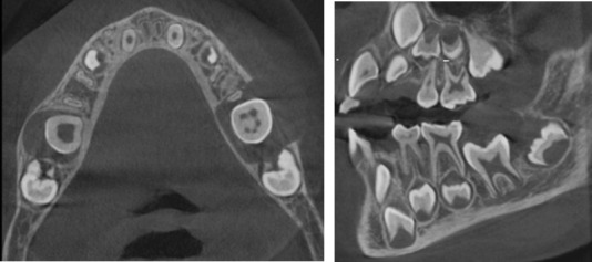
Dentigerous cysts are a common cystic pathology that develop between the first and third decade of life and are mainly associated with impacted or erupted mandibular third molars followed by maxillary canines and maxillary third molars. These kinds of cysts are the result of the proliferation of enamel epithelium after its formation, the pathogenesis of which is not clear. Few of these cysts have been reported in pediatric patients. The following case report presents the rare occurrence of a dentigerous cyst in a 6-year-old boy and describes the treatment administered.
Los quistes dentígeros son una patología quística común que se desarrolla entre la primera y la tercera década de la vida, y se asocian principalmente con terceros molares mandibulares incluidos o erupcionados, seguidos de caninos superiores y terceros molares superiores. Este tipo de quistes son el resultado de la proliferación del epitelio del esmalte después de su formación, cuya patogenia no está clara. Se han informado pocos de estos quistes en pacientes pediátricos. El siguiente reporte de caso presenta la rara ocurrencia de un quiste dentígero en un niño de 6 años y describe el tratamiento administrado.
Keywords: cone beam computed tomography; dentigerous cyst; developmental cyst; pediatric.
Conflict of interest statement
Conflict of interests: The authors do not report any conflict of interest
Figures


Similar articles
-
Dentigerous cysts in four quadrants: a rare and first reported case.J Surg Tech Case Rep. 2013 Jan;5(1):21-6. doi: 10.4103/2006-8808.118607. J Surg Tech Case Rep. 2013. PMID: 24470846 Free PMC article.
-
Anterior maxillary dentigerous cyst.Int J Clin Pediatr Dent. 2009 Jan;2(1):42-5. doi: 10.5005/jp-journals-10005-1041. Epub 2009 Apr 26. Int J Clin Pediatr Dent. 2009. PMID: 25206099 Free PMC article.
-
Bi-maxillary dentigerous cyst in a non-syndromic child - review of literature with a case presentation.J Stomatol Oral Maxillofac Surg. 2017 Feb;118(1):45-48. doi: 10.1016/j.jormas.2016.12.001. Epub 2017 Feb 3. J Stomatol Oral Maxillofac Surg. 2017. PMID: 28330574 Review.
-
A Case Series of Dentigerous Cyst in Paediatric Patients at Our Tertiary Institution.Indian J Otolaryngol Head Neck Surg. 2023 Sep;75(3):2444-2452. doi: 10.1007/s12070-023-03782-6. Epub 2023 Apr 18. Indian J Otolaryngol Head Neck Surg. 2023. PMID: 37636721 Free PMC article.
-
Large pediatric maxillary dentigerous cysts presenting with sinonasal and orbital symptoms: A case series.Ear Nose Throat J. 2017 Apr-May;96(4-5):E29-E34. Ear Nose Throat J. 2017. PMID: 28489242 Review.
References
Publication types
LinkOut - more resources
Full Text Sources