Electron Beam Powder Bed Fusion of Ti-48Al-2Cr-2Nb Open Porous Scaffold for Biomedical Applications: Process Parameters, Adhesion, and Proliferation of NIH-3T3 Cells
- PMID: 38389689
- PMCID: PMC10880641
- DOI: 10.1089/3dp.2022.0108
Electron Beam Powder Bed Fusion of Ti-48Al-2Cr-2Nb Open Porous Scaffold for Biomedical Applications: Process Parameters, Adhesion, and Proliferation of NIH-3T3 Cells
Abstract
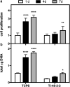
Titanium aluminide (TiAl)-based intermetallics, especially Ti-48Al-2Cr-2Nb, are a well-established class of materials for producing bulky components using the electron beam powder bed fusion (EB-PBF) process. The biological properties of Ti-48Al-2Cr-2Nb alloy have been rarely investigated, specifically using complex cellular structures. This work investigates the viability and proliferation of NIH-3T3 fibroblasts on Ti-48Al-2Cr-2Nb dodecahedral open scaffolds manufactured by the EB-PBF process. A process parameter optimization is carried out to produce a fully dense part. Then scaffolds are produced and characterized using different techniques, including scanning electron microscopy and X-ray tomography. In vitro viability tests are performed with NIH-3T3 cells after incubation for 1, 4, and 7 days. The results show that Ti-48Al-2Cr-2Nb represents a promising new entry in the biomaterial field.
Keywords: 3D printing; X-ray analysis; additive manufacturing; biocompatibility; titanium aluminide.
Copyright 2024, Mary Ann Liebert, Inc., publishers.
Conflict of interest statement
No competing financial interests exist.
Figures


Similar articles
-
Additive Manufacturing of Ti-48Al-2Cr-2Nb Alloy Using Gas Atomized and Mechanically Alloyed Plasma Spheroidized Powders.Materials (Basel). 2020 Sep 7;13(18):3952. doi: 10.3390/ma13183952. Materials (Basel). 2020. PMID: 32906691 Free PMC article.
-
Corrosion evaluation of Ti-48Al-2Cr-2Nb (at.%) in Ringer's solution.Acta Biomater. 2006 Nov;2(6):701-8. doi: 10.1016/j.actbio.2006.05.012. Epub 2006 Aug 2. Acta Biomater. 2006. PMID: 16887397
-
In vitro evaluation of human osteoblast adhesion to a thermally oxidized gamma-TiAl intermetallic alloy of composition Ti-48Al-2Cr-2Nb (at.%).J Mater Sci Mater Med. 2010 May;21(5):1739-50. doi: 10.1007/s10856-010-4016-6. Epub 2010 Feb 17. J Mater Sci Mater Med. 2010. PMID: 20162332 Free PMC article.
-
Powder based additive manufacturing for biomedical application of titanium and its alloys: a review.Biomed Eng Lett. 2020 Oct 26;10(4):505-516. doi: 10.1007/s13534-020-00177-2. eCollection 2020 Nov. Biomed Eng Lett. 2020. PMID: 33194244 Free PMC article. Review.
-
Advancements in Additive Manufacturing of Tantalum via the Laser Powder Bed Fusion (PBF-LB/M): A Comprehensive Review.Materials (Basel). 2023 Sep 27;16(19):6419. doi: 10.3390/ma16196419. Materials (Basel). 2023. PMID: 37834556 Free PMC article. Review.
References
-
- Galati M, Iuliano L. A literature review of powder-based electron beam melting focusing on numerical simulations. Addit Manuf 2018;19:1–20; doi: 10.1016/j.addma.2017.11.001 - DOI
-
- Saboori A, Abdi A, Fatemi SA, et al. . Hot deformation behavior and flow stress modeling of Ti–6Al–4V alloy produced via electron beam melting additive manufacturing technology in single β-phase field. Mater Sci Eng A 2020;792:139822; doi: 10.1016/j.msea.2020.139822 - DOI
-
- Del Guercio G, Galati M, Saboori A, et al. . Microstructure and mechanical performance of Ti-6Al-4V lattice structures manufactured via electron beam melting (EBM): A review. Acta Metall Sin (English Lett) 2020;33(2):183–203; doi: 10.1007/s40195-020-00998-1 - DOI
-
- Loeber L, Biamino S, Ackelid U, et al. . Comparison of selective laser and electron beam melted titanium aluminides. In: 22nd Annual International Solid Freeform Fabrication Symposium—An Additive Manufacturing Conference, SFF. Austin, Texas, USA, 2011.
LinkOut - more resources
Full Text Sources
