. 2023 Dec 21;15(2):704-719.
doi: 10.1039/d3md00630a. eCollection 2024 Feb 21.

Highly potent and selective phosphatidylinositol 4-kinase IIIβ inhibitors as broad-spectrum anti-rhinoviral agents

Affiliations

Highly potent and selective phosphatidylinositol 4-kinase IIIβ inhibitors as broad-spectrum anti-rhinoviral agents

Avinash G Vishakantegowda et al. RSC Med Chem. .

Erratum in

Abstract

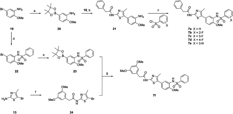
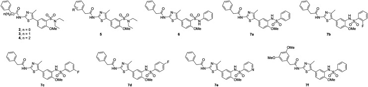
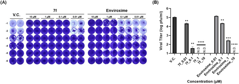
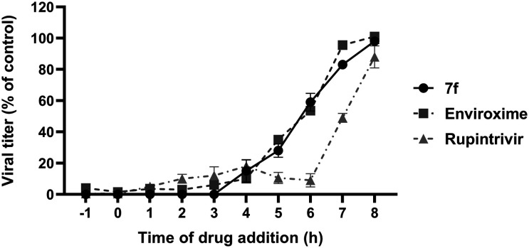
Human rhinoviruses (hRVs) cause upper and lower respiratory tract infections and exacerbate asthma and chronic obstructive pulmonary disease. hRVs comprise more than 160 strains with considerable genetic variation. Their high diversity and strain-specific interactions with antisera hinder the development of anti-hRV therapeutic agents. Phosphatidylinositol-4-kinase IIIβ (PI4KIIIβ) is a key enzyme in the phosphoinositide signalling pathway that is crucial for the replication and survival of various viruses. We identified novel PI4KIIIβ inhibitors, N-(4-methyl-5-arylthiazol)-2-amide derivatives, by generating a hit compound, 1a, from the high-throughput screening of a chemical library, followed by the optimization study of 1a. Inhibitor 7e exhibited the highest activity (EC50 = 0.008, 0.0068, and 0.0076 μM for hRV-B14, hRV-A16, and hRV-A21, respectively) and high toxicity (CC50 = 6.1 μM). Inhibitor 7f showed good activity and low toxicity and provided the highest selectivity index (SI ≥ 4638, >3116, and >2793 for hRV-B14, hRV-A16, and hRV-A21, respectively). Furthermore, 7f showed broad-spectrum activities against various hRVs, coxsackieviruses, and other enteroviruses, such as EV-A71 and EV-D68. The binding mode of the inhibitors was investigated using 7f, and the experimental results of plaque reduction, replicon and cytotoxicity, and time-of-drug-addition assays suggested that 7f acts as a PI4KIIIβ inhibitor. The kinase inhibition activity of this series of compounds against PI4KIIIα and PI4KIIIβ was assessed, and 7f demonstrated kinase inhibition activity with an IC50 value of 0.016 μM for PI4KIIIβ, but not for PI4KIIIα (>10 μM). Therefore, 7f represents a highly potent and selective PI4KIIIβ inhibitor for the further development of antiviral therapy against hRVs or other enteroviruses.


Conflict of interest statement

There are no conflicts to declare.

Figures

Fig. 1
Fig. 1. (A) Structure of known PI4KIIIβ inhibitors and (B) core structures of new N-[5-(3-sulfamoyl-4-methoxyphenyl)-4-methylthiazol-2-yl]-2-phenylacetamide and its derivatives 5–7.
Scheme 1
Scheme 1. Synthesis of compounds 2–4 and 5a–5l. 5l reagents and conditions: (a) chlorosulfonic acid, 0 °C to rt, 50%; (b) diethylamine, DIPEA, THF, 0 °C to rt, 85%; (c) bis(pinacolato)diboron, PdCl2(dppf)·DCM, potassium acetate, 1,4-dioxane, 90 °C, 46%; (d) sulfuric acid (20% aq.), bromine, 0 °C to rt, 80%; (e) trifluoroacetic anhydride, 16 TEA, THF, 0 °C to rt, 72%; (f) Pd(PPh3)4, tripotassium phosphate, 1,4-dioxane/water, 90 °C, 62%; (g) RCO2H, HATU, DIPEA, DCM, rt, 9–90%.
Scheme 2
Scheme 2. Synthesis of compound 6. Reagents and conditions: (a) aniline, DIPEA, THF, 0 °C to rt, 97%; (b) bis(pinacolato)diboron, PdCl2(dppf)·DCM, potassium acetate, 1,4-dioxane, 90 °C, 35%; (c) phenylacetic acid, HATU, DIPEA, DCM, rt, 33%; (d) Pd(PPh3)4, tripotassium phosphate, 1,4-dioxane/water, 90 °C, 46%.
Scheme 3
Scheme 3. Synthesis of compounds 7a–7f. Reagents and conditions: (a) bis(pinacolato)diboron, PdCl2(dppf)·DCM, potassium acetate, DMSO, 80 °C, 65%; (b) Pd(PPh3)4, 0.5 M K2CO3 aq., 1,4-dioxane, 90 °C, 37%; (c) aryl sulfonyl chloride, pyridine, rt, 32–82%; (d) phenyl sulfonyl chloride, pyridine, rt, 99%; (e) bis(pinacolato)diboron, PdCl2(dppf)·DCM, potassium acetate, 1,4-dioxane, 90 °C, 53%; (f) 3,5-dimethoxy phenylacetic acid, HATU, DIPEA, DCM, rt, 53%; (g) Pd(PPh3)4, tripotassium phosphate, 1,4-dioxane/water, 90 °C, 47%.
Fig. 2
Fig. 2. Structures of newly synthesized N-[5-(3-sulfamoyl-4-methoxyphenyl)-4-methylthiazol-2-yl]-2-phenylacetamide and its derivatives 2–7.
Fig. 3
Fig. 3. Plaque reduction assay. (A) H1HeLa cells were infected with dose-dependent 7f and enviroxime and incubated for 3 days. Plaques were visualized by crystal violet staining. (B) Dose–response curve analysis of hRV14 inhibition by 7f and enviroxime. Data represent means (±SD) of at least two dependent experiments performed in duplicate. Significant differences are indicated by *p < 0.05, **p < 0.01, ***p < 0.001, ****p < 0.0001.
Fig. 4
Fig. 4. Replicon and cytotoxicity assay. (A and B) H1HeLa cells were transfected with the RNA transcripts of the EV71 replicon and immediately treated with each serially diluted compound for 8 h. Dose–response inhibition rate (black bar) and cytotoxicity (open square) in the presence of serially diluted concentrations of 7f (A) and enviroxime (B). Data represent means (±SD) of at least two dependent experiments performed in duplicate. Significant differences are indicated by *p < 0.05, **p < 0.01, ***p < 0.001, ****p < 0.0001.
Fig. 5
Fig. 5. Time-of-drug-addition assay. hRV14 infected H1HeLa cells were treated with 7f (1 μM), enviroxime (5 μM), and rupintrivir (0.5 μM) at 1h intervals. At 9 h post-infection, cell lysates were harvested, and the virus titre was determined by endpoint titration. The relative viral titres of 7f (black circle), enviroxime (grey square), and rupintrivir (grey triangle) were calculated as a percentage of the virus control. Data represent means (±SD) of at least two dependent experiments performed in duplicate.
Fig. 6
Fig. 6. Docking model of 7f (pink ball and stick model) with PI4KIIIβ (blue ribbon model). The hydrogen bonds are displayed as green dashed lines, and the hydrophobic interactions are shown as pink dashed lines. The key interaction residues are visible in the stick model and are labelled using the 3-letter amino acid code.

References

    1. Aab A. Wirz O. van de Veen W. Sollner S. Stanic B. Ruckert B. Aniscenko J. Edwards M. R. Johnston S. L. Papadopoulos N. G. Rebane A. Akdis C. A. Akdis M. Allergy. 2017;72:232–243. doi: 10.1111/all.12931. - DOI - PubMed
    1. Gern J. E. Clin. Exp. Allergy. 2015;45:32–42. doi: 10.1111/cea.12428. - DOI - PubMed
    1. Message S. D. Laza-Stanca V. Mallia P. Parker H. L. Zhu J. Kebadze T. Contoli M. Sanderson G. Kon O. M. Papi A. Jeffery P. K. Stanciu L. A. Johnston S. L. Proc. Natl. Acad. Sci. U. S. A. 2008;105:13562–13567. - PMC - PubMed
    1. Papadopoulos N. G. Sanderson G. Hunter J. Johnston S. L. J. Med. Virol. 1999;58:100–104. doi: 10.1002/(SICI)1096-9071(199905)58:1<100::AID-JMV16>3.0.CO;2-D. - DOI - PubMed
    1. Patick A. K. Binford S. L. Brothers M. A. Jackson R. L. Ford C. E. Diem M. D. Maldonado F. Dragovich P. S. Zhou R. Prins T. J. Fuhrman S. A. Meador J. W. Zalman L. S. Matthews D. A. Worland S. T. Antimicrob. Agents Chemother. 1999;43:2444–2450. - PMC - PubMed

LinkOut - more resources