

The phenomenon of phototoxicity and long-term risks of commonly prescribed and structurally diverse drugs
- PMID: 38389933
- PMCID: PMC10883358
- DOI: 10.1016/j.jpap.2023.100221
The phenomenon of phototoxicity and long-term risks of commonly prescribed and structurally diverse drugs
Abstract
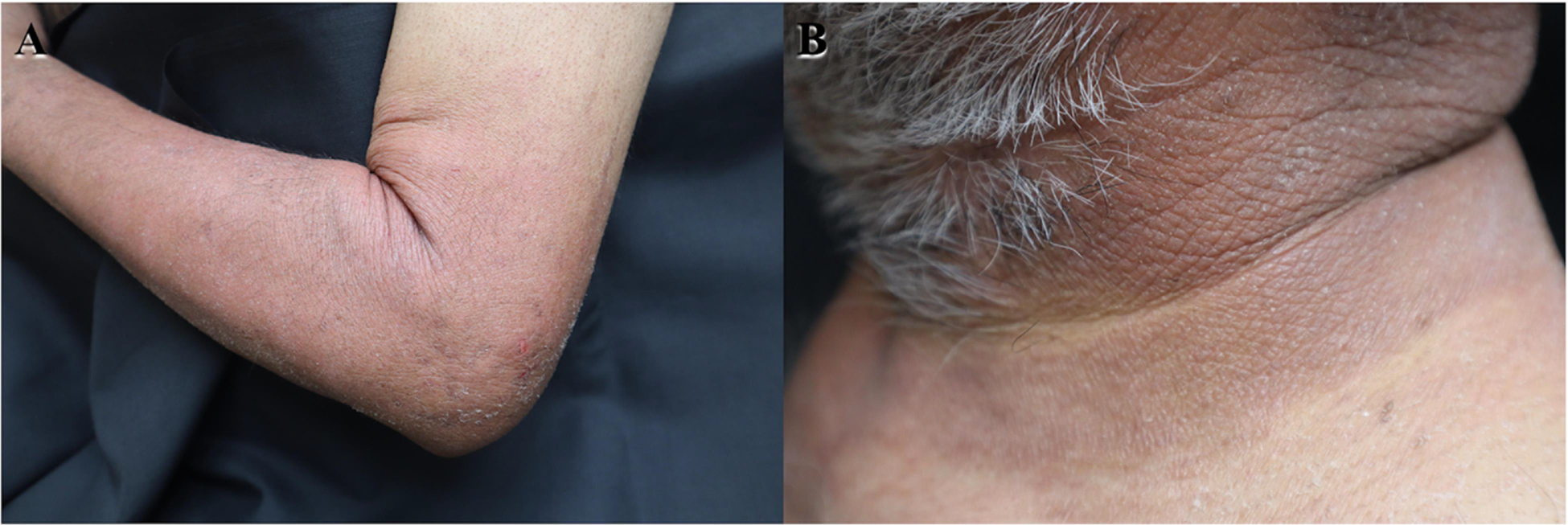
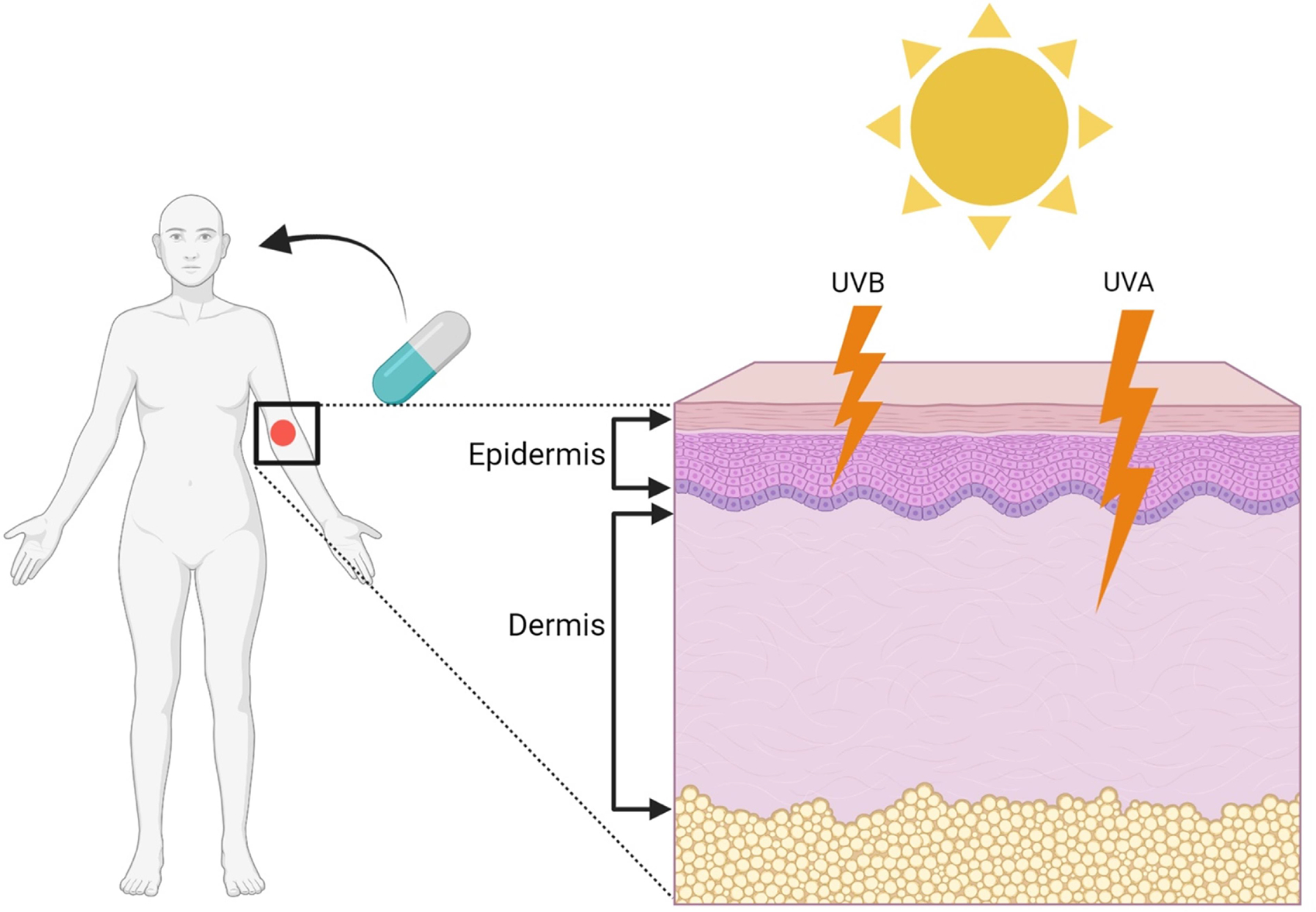
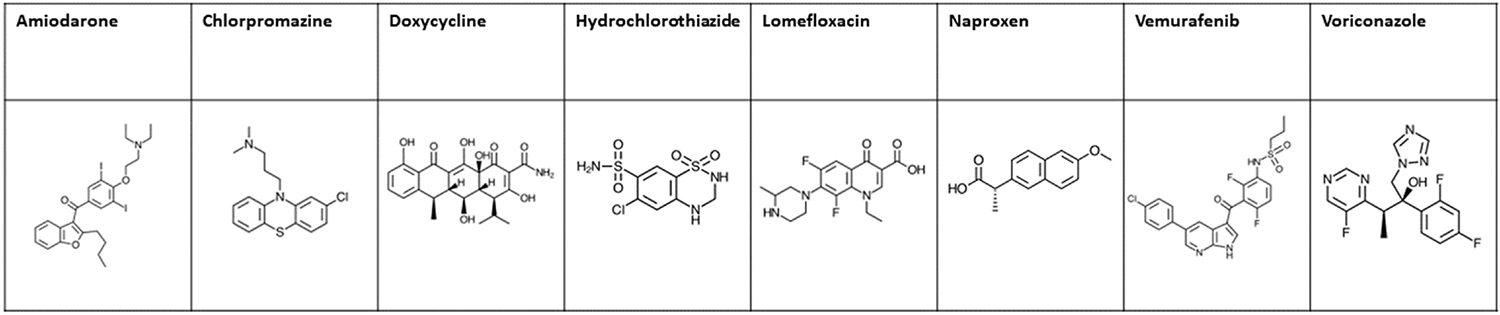
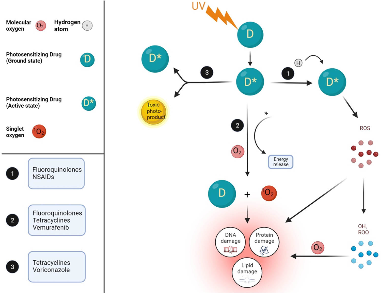
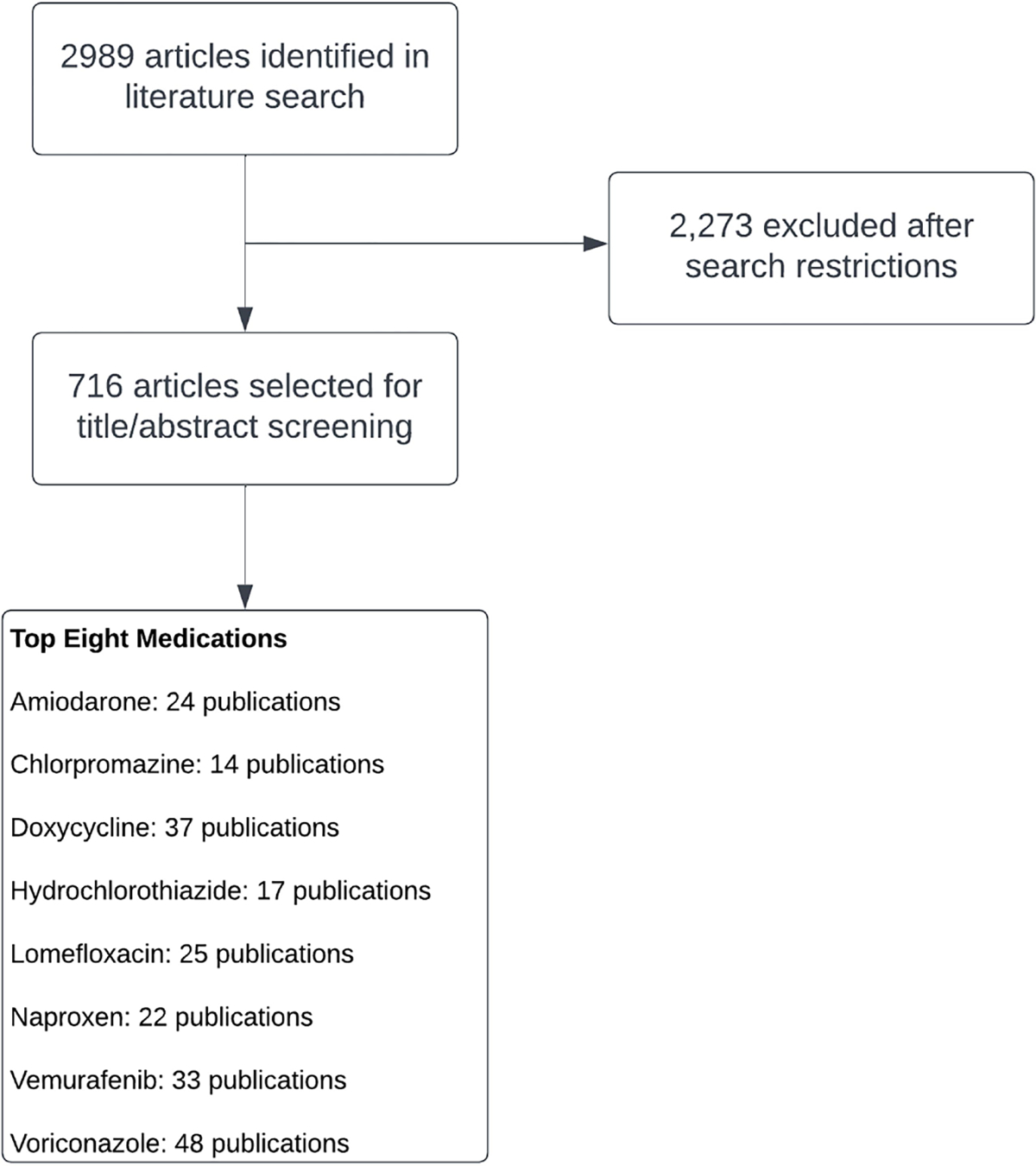
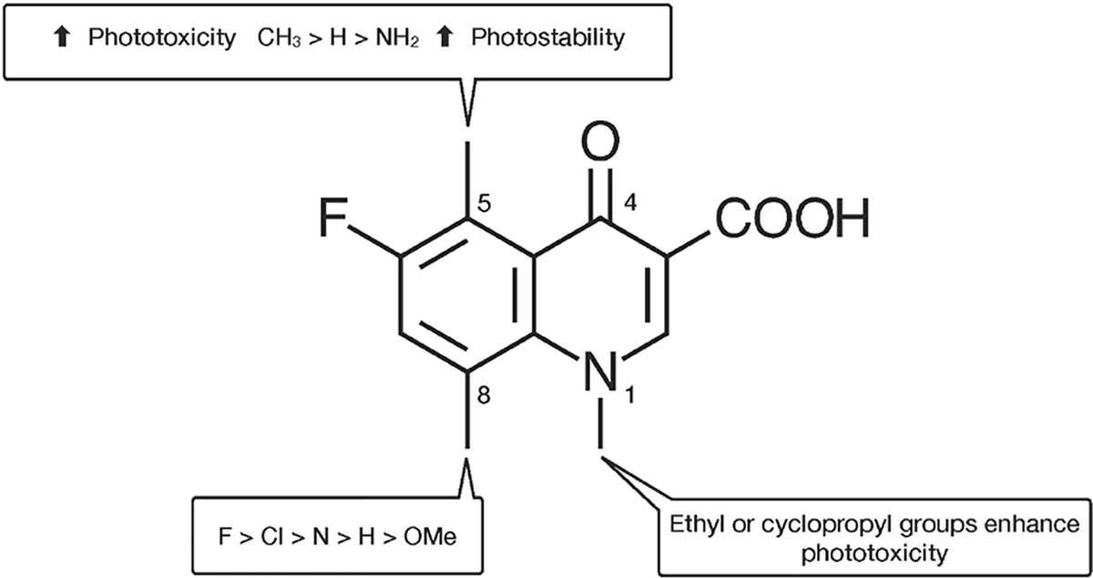
Photosensitivity to structurally diverse drugs is a common but under-reported adverse cutaneous reaction and can be classified as phototoxic or photoallergic. Phototoxic reactions occur when the skin is exposed to sunlight after administering topical or systemic medications that exhibit photosensitizing activity. These reactions depend on the dose of medication, degree of exposure to ultraviolet light, type of ultraviolet light, and sufficient skin distribution volume. Accurate prediction of the incidence and phototoxic response severity is challenging due to a paucity of literature, suggesting that phototoxicity may be more frequent than reported. This paper reports an extensive literature review on phototoxic drugs; the review employed pre-determined search criteria that included meta-analyses, systematic reviews, literature reviews, and case reports freely available in full text. Additional reports were identified from reference sections that contributed to the understanding of phototoxicity. The following drugs and/or drug classes are discussed: amiodarone, voriconazole, chlorpromazine, doxycycline, fluoroquinolones, hydrochlorothiazide, nonsteroidal anti-inflammatory drugs, and vemurafenib. In reviewing phototoxic skin reactions, this review highlights drug molecular structures, their reactive pathways, and, as there is a growing association between photosensitizing drugs and the increasing incidence of skin cancer, the consequential long-term implications of photocarcinogenesis.
Keywords: Drug reaction; Photosensitizing medications; Phototoxicity; Reactive oxygen species; Skin cancer; Ultraviolet light.
Figures
Similar articles
-
Photosensitizing drug reactions.Clin Dermatol. 2022 Jan-Feb;40(1):57-63. doi: 10.1016/j.clindermatol.2021.08.014. Epub 2021 Aug 8. Clin Dermatol. 2022. PMID: 35190066 Review.
-
Phototoxic and photoallergic cutaneous drug reactions.Chem Immunol Allergy. 2012;97:167-79. doi: 10.1159/000335630. Epub 2012 May 3. Chem Immunol Allergy. 2012. PMID: 22613861
-
Drug-induced phototoxicity: A systematic review.J Am Acad Dermatol. 2018 Dec;79(6):1069-1075. doi: 10.1016/j.jaad.2018.06.061. Epub 2018 Jul 10. J Am Acad Dermatol. 2018. PMID: 30003982
-
Knowledge about Ultraviolet Radiation Hazards and Tanning Behavior of Cosmetology and Medical Students.Acta Dermatovenerol Croat. 2016 Apr;24(1):73-7. Acta Dermatovenerol Croat. 2016. PMID: 27149135
-
Photosensitivity to exogenous agents.J Cutan Med Surg. 2004 Nov-Dec;8(6):424-31. doi: 10.1007/s10227-005-0017-3. J Cutan Med Surg. 2004. PMID: 15988550 Review.
Cited by
-
The Importance of Murine Models in Determining In Vivo Pharmacokinetics, Safety, and Efficacy in Antimalarial Drug Discovery.Pharmaceuticals (Basel). 2025 Mar 18;18(3):424. doi: 10.3390/ph18030424. Pharmaceuticals (Basel). 2025. PMID: 40143200 Free PMC article. Review.
-
Drugs Associated with Adverse Effects in Vulnerable Groups of Patients.Clin Pract. 2024 May 31;14(3):1010-1020. doi: 10.3390/clinpract14030080. Clin Pract. 2024. PMID: 38921258 Free PMC article. Review.
-
Causes of drug-induced photosensitivity: an analysis using FDA adverse event reporting system database.Sci Rep. 2025 May 24;15(1):18102. doi: 10.1038/s41598-025-03114-4. Sci Rep. 2025. PMID: 40413262 Free PMC article.
-
Current Insights into the Role of UV Radiation-Induced Oxidative Stress in Melanoma Pathogenesis.Int J Mol Sci. 2024 Oct 30;25(21):11651. doi: 10.3390/ijms252111651. Int J Mol Sci. 2024. PMID: 39519202 Free PMC article. Review.
References
-
- Selvaag E, Clinical drug photosensitivity. A retrospective analysis of reports to the Norwegian adverse drug reactions committee from the years 1970–1994, Photodermatol. Photoimmunol. Photomed. 13 (1–2) (1997) 21–23. - PubMed
-
- Chaabane H, Masmoudi A, Amouri M, Ghorbel S, Boudaya S, Hammami S, et al., [Cutaneous adverse drug reaction: prospective study of 118 cases], Tunis Med. 91 (8–9) (2013) 514–520. - PubMed
-
- James WE, Treat DM, Rosenbach JR, Neuhaus MA, Elsevier, IM. dermatoses resulting from physical factors, editor. Andrews’ Diseases of the Skin: Clinical Dermatology, 13, Elsevier, Philadelphia, PA, 2020, pp. 18–45.
-
- Kerr HA, Lim HW, Photodermatoses in African Americans: a retrospective analysis of 135 patients over a 7-year period, J. Am. Acad. Dermatol. 57 (4) (2007) 638–643. - PubMed
-
- Nakamura M, Henderson M, Jacobsen G, Lim HW, Comparison of photodermatoses in African-Americans and Caucasians: a follow-up study, Photodermatol. Photoimmunol. Photomed. 30 (5) (2014) 231–236. - PubMed
Grants and funding
LinkOut - more resources
Full Text Sources