Review
. 2024 Feb 7;11(1):79-100.
doi: 10.3390/dermatopathology11010008.

Galli-Galli Disease: A Comprehensive Literature Review

Affiliations
Review

Galli-Galli Disease: A Comprehensive Literature Review

Andrea Michelerio et al. Dermatopathology (Basel). .

Abstract

Galli-Galli disease (GGD) is a rare genodermatosis that exhibits autosomal dominant inheritance with variable penetrance. GGD typically manifests with erythematous macules, papules, and reticulate hyperpigmentation in flexural areas. A distinct atypical variant exists, which features brown macules predominantly on the trunk, lower limbs, and extremities, with a notable absence of the hallmark reticulated hyperpigmentation in flexural areas. This review includes a detailed literature search and examines cases since GGD's first description in 1982. It aims to synthesize the current knowledge on GGD, covering its etiology, clinical presentation, histopathology, diagnosis, and treatment. A significant aspect of this review is the exploration of the genetic, histopathological, and clinical parallels between GGD and Dowling-Degos disease (DDD), which is another rare autosomal dominant genodermatosis, particularly focusing on their shared mutations in the KRT5 and POGLUT1 genes. This supports the hypothesis that GGD and DDD may be different phenotypic expressions of the same pathological condition, although they have traditionally been recognized as separate entities, with suprabasal acantholysis being a distinctive feature of GGD. Lastly, this review discusses the existing treatment approaches, underscoring the absence of established guidelines and the limited effectiveness of various treatments.

Keywords: acantholysis; genetic; hyperpigmentation; keratin-5; papulosquamous; skin diseases.


Conflict of interest statement

The authors declare no conflicts of interest.

Figures

Figure 1
Figure 1
Schematic diagram of the dermal–epidermal junction.
Figure 2
Figure 2
Overview of Notch signaling. Notch receptors undergo initial translation in the endoplasmic reticulum and are then trafficked to the Golgi apparatus. During trafficking, O-linked and N-linked glycosylations occur. Then, in the Golgi apparatus, Notch receptors are cleaved into heterodimers (S1 cleavage) and transported to the cell membrane. Upon reaching the cell surface, the receptor is activated through ligand binding. This ligand–receptor interaction triggers transendocytosis in the neighboring cell, inducing a conformational alteration in the Notch receptor and exposing the S2 site. This site is cleaved by ADAM metalloproteases (S2 cleavage), generating a membrane-anchored Notch extracellular truncation (NEXT). The S2 cleavage exposes S3 and S4 sites, facilitating further proteolytic cleavage by the γ-secretase complex (S3/S4 cleavage). This results in the liberation of the notch intracellular domain (NICD), which translocates to the nucleus. There, it binds to CBF1/RBP-Jκ/Su(H)/Lag-1 (CSL), which is known as RBP-Jκ in vertebrates. In its basal state, CSL functions as a transcriptional repressor by associating with corepressor (Co-R) proteins. NICD binding converts it into a transcriptional activator by forming a ternary complex with mastermind-like (MAML) proteins, thus initiating the transcription of downstream target genes [29].
Figure 3
Figure 3
Typical presentation of GGD with hyperkeratotic papules and erythematous brownish macules that coalesce into patches with a reticulated appearance on the inguinal (A) and axillary folds (B).
Figure 4
Figure 4
Atypical presentation of GGD with dissemination of brownish macules and erythematous papules on the trunk with sparing of the folds (A,B).
Figure 5
Figure 5
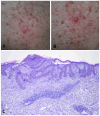
During flare phases of GGD, multiple vesicles develop, leading to intensely pruritic erosions and crusts on the skin (A,B). Histopathology shows a superficial dermal perivascular lymphocytic infiltrate. Apical acantholysis is apparent in the epidermis (C).
Figure 6
Figure 6
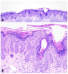
During the noninflammatory phase of GGD, the dermatosis is histologically indistinguishable from DDD. The epidermis shows thin, elongated, branching rete ridges with basal hyperpigmentation. There is mild hyperkeratosis overlying the epidermis (A,B).

References

    1. Gilchrist H., Jackson S., Morse L., Nesbitt L.T. Galli-Galli disease: A case report with review of the literature. J. Am. Acad. Dermatol. 2008;58:299–302. doi: 10.1016/j.jaad.2007.05.041. - DOI - PubMed
    1. Braun-Falco M., Volgger W., Borelli S., Ring J., Disch R. Galli-Galli disease: An unrecognized entity or an acantholytic variant of Dowling-Degos disease? J. Am. Acad. Dermatol. 2001;45:760–763. doi: 10.1067/mjd.2001.116340. - DOI - PubMed
    1. Bardach H., Gebhart W., Luger T. Genodermatose bei einem Brüderpaar: Morbus Dowling-Degos, Grover, Darier, Hailey-Hailey oder Galli-Galli? [Genodermatosis in a pair of brothers: Dowling-Degos, Grover, Darier, Hailey-Hailey or Galli-Galli disease?] Hautarzt. 1982;33:378–383. - PubMed
    1. Rundle C.W., Ophaug S., Simpson E.L. Acitretin therapy for Galli-Galli disease. JAAD Case Rep. 2020;6:457–461. doi: 10.1016/j.jdcr.2020.02.042. - DOI - PMC - PubMed
    1. Yang A., Cheung K., Kossard S., Murrell D.F. Atypical Disseminated Variant of Galli–Galli Disease: A Review of the Literature. Am. J. Dermatopathol. 2020;42:484–490. doi: 10.1097/DAD.0000000000001467. - DOI - PubMed