

Left ventricular hypertrophy as a risk factor for accelerated brain aging: Results from the Study of Health in Pomerania
- PMID: 38391110
- PMCID: PMC10885183
- DOI: 10.1002/hbm.26567
Left ventricular hypertrophy as a risk factor for accelerated brain aging: Results from the Study of Health in Pomerania
Abstract
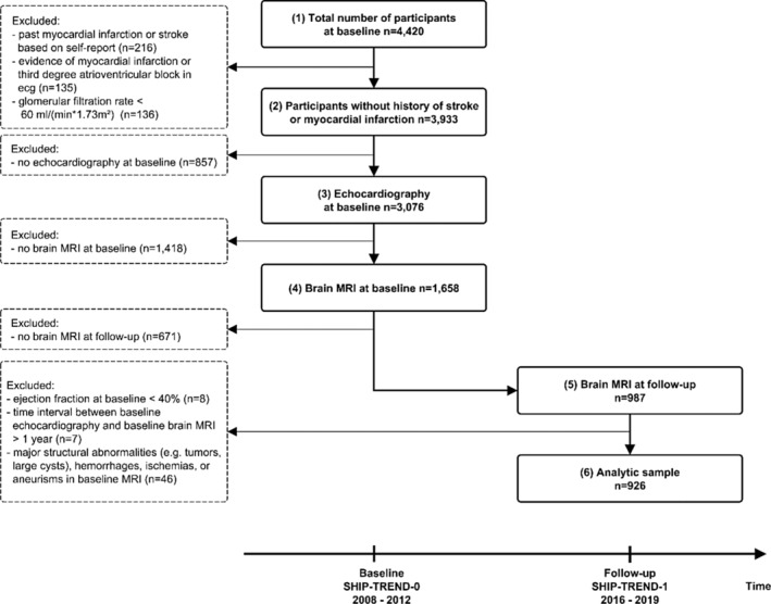
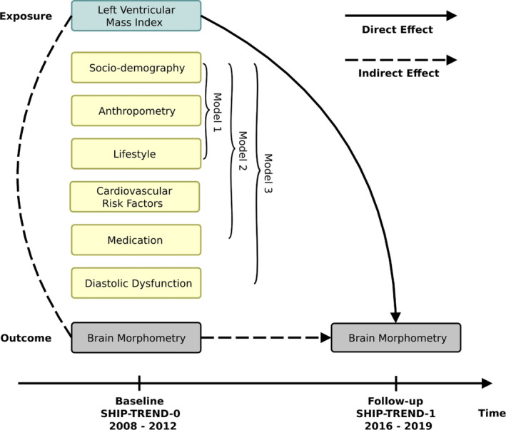
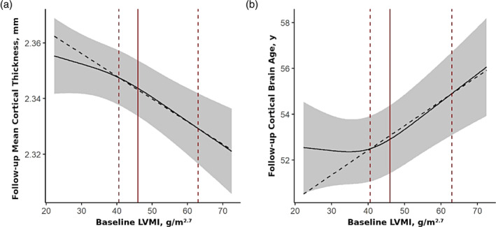
Previous studies provided evidence for the importance of cardiac structure abnormalities, in particular greater left ventricular (LV) mass, for brain aging, but longitudinal studies are lacking to date. We included 926 individuals (median age 48 years; 53% women) from the TREND cohort of the Study of Health in Pomerania (SHIP) without reduced ejection fraction or a history of myocardial infarction. LV mass index (LVMI) was determined by echocardiography at baseline. Brain morphometric measurements were derived from magnetic resonance images at baseline and 7-year follow-up. Direct effects of baseline LVMI on brain morphometry at follow-up were estimated using linear regression models with adjustment for baseline brain morphometry. At baseline, median LVMI was 40 g/m2.7 and 241 individuals (26%) met the criterion of LV hypertrophy. After correction for multiple testing, baseline LVMI was directly associated with reduced global cortical thickness and increased cortical brain age at follow-up independent from hypertension and blood pressure. Exposure-outcome relations were nonlinear and significantly stronger in the upper half of the exposure distribution. Specifically, an increase in baseline LVMI from the 50% quantile to the 95% quantile was associated additional 2.7 years (95% confidence interval = [1.5 years, 3.8 years]) of cortical brain age at follow-up. Additional regional analyses yielded bilateral effects on multiple frontal cortical regions. Our findings highlight the role of cardiac structure in brain aging. LVMI constitutes an easily measurable marker that might help to identify persons at risk for cognitive impairment and dementia.
Keywords: brain aging; brain imaging; epidemiology; left ventricular hypertrophy.
© 2024 The Authors. Human Brain Mapping published by Wiley Periodicals LLC.
Conflict of interest statement
HJG has received travel grants and speakers honoraria from Fresenius Medical Care, Neuraxpharm, Servier, and Janssen Cilag as well as research funding from Fresenius Medical Care. All other authors have nothing to disclose.
Figures
Similar articles
-
Cardiac Hypertrophy Is Associated With Advanced Brain Aging in the General Population.J Am Heart Assoc. 2021 Sep 7;10(17):e020994. doi: 10.1161/JAHA.121.020994. Epub 2021 Sep 1. J Am Heart Assoc. 2021. PMID: 34465186 Free PMC article.
-
Prognostic implications of left ventricular mass and geometry following myocardial infarction: the VALIANT (VALsartan In Acute myocardial iNfarcTion) Echocardiographic Study.JACC Cardiovasc Imaging. 2008 Sep;1(5):582-91. doi: 10.1016/j.jcmg.2008.05.012. JACC Cardiovasc Imaging. 2008. PMID: 19356485 Clinical Trial.
-
Ambulatory systolic blood pressure and obesity are independently associated with left ventricular hypertrophic remodeling in children.J Cardiovasc Magn Reson. 2017 Nov 9;19(1):86. doi: 10.1186/s12968-017-0401-3. J Cardiovasc Magn Reson. 2017. PMID: 29117866 Free PMC article.
-
A History of Asthma From Childhood and Left Ventricular Mass in Asymptomatic Young Adults: The Bogalusa Heart Study.JACC Heart Fail. 2017 Jul;5(7):497-504. doi: 10.1016/j.jchf.2017.03.009. Epub 2017 Jun 26. JACC Heart Fail. 2017. PMID: 28662937 Free PMC article.
-
Assessment of prevalence of left ventricular hypertrophy in hypertension.J Hypertens. 1998 Jun;16(6):715-23. doi: 10.1097/00004872-199816060-00001. J Hypertens. 1998. PMID: 9663910 Review.
Cited by
-
Cognitive Outcomes in Young Adults with Primary Arterial Hypertension: The Role of Cardiovascular Risk Factors and Hypertension-Mediated Organ Damage.Medicina (Kaunas). 2024 Aug 20;60(8):1353. doi: 10.3390/medicina60081353. Medicina (Kaunas). 2024. PMID: 39202634 Free PMC article.
-
Progress in diagnosis and treatment of hypertension combined with left ventricular hypertrophy.Ann Med. 2024 Dec;56(1):2405080. doi: 10.1080/07853890.2024.2405080. Epub 2024 Sep 20. Ann Med. 2024. PMID: 39301864 Free PMC article. Review.
References
-
- Bluemke, D. A. , Kronmal, R. A. , Lima, J. A. C. , Liu, K. , Olson, J. , Burke, G. L. , & Folsom, A. R. (2008). The relationship of left ventricular mass and geometry to incident cardiovascular events. Journal of the American College of Cardiology, 52(25), 2148–2155. 10.1016/j.jacc.2008.09.014 - DOI - PMC - PubMed
-
- Bülow, R. , Ittermann, T. , Dörr, M. , Poesch, A. , Langner, S. , Völzke, H. , Hosten, N. , & Dewey, M. (2018). Reference ranges of left ventricular structure and function assessed by contrast‐enhanced cardiac MR and changes related to ageing and hypertension in a population‐based study. European Radiology, 28(9), 3996–4005. 10.1007/s00330-018-5345-y - DOI - PubMed
-
- Cermakova, P. , Muller, M. , Armstrong, A. C. , Religa, D. , Bryan, R. N. , Lima, J. A. C. , & Launer, L. J. (2017). Subclinical cardiac dysfunction and brain health in midlife: CARDIA (coronary artery risk development in young adults) brain magnetic resonance imaging substudy. Journal of the American Heart Association, 6(12), e006750. 10.1161/JAHA.117.006750 - DOI - PMC - PubMed
-
- Cole, J. H. , Ritchie, S. J. , Bastin, M. E. , Valdés Hernández, M. C. , Muñoz Maniega, S. , Royle, N. , Corley, J. , Pattie, A. , Harris, S. E. , Zhang, Q. , Wray, N. R. , Redmond, P. , Marioni, R. E. , Starr, J. M. , Cox, S. R. , Wardlaw, J. M. , Sharp, D. J. , & Deary, I. J. (2018). Brain age predicts mortality. Molecular Psychiatry, 23(5), 1385–1392. 10.1038/mp.2017.62 - DOI - PMC - PubMed
MeSH terms
Grants and funding
LinkOut - more resources
Full Text Sources
Medical
Miscellaneous
