Review
. 2024 Jan 23;11(2):104.
doi: 10.3390/bioengineering11020104.

Label-Free Optical Technologies for Middle-Ear Diseases

Affiliations
Review

Label-Free Optical Technologies for Middle-Ear Diseases

Zeyi Zhou et al. Bioengineering (Basel). .

Abstract

Medical applications of optical technology have increased tremendously in recent decades. Label-free techniques have the unique advantage of investigating biological samples in vivo without introducing exogenous agents. This is especially beneficial for a rapid clinical translation as it reduces the need for toxicity studies and regulatory approval for exogenous labels. Emerging applications have utilized label-free optical technology for screening, diagnosis, and surgical guidance. Advancements in detection technology and rapid improvements in artificial intelligence have expedited the clinical implementation of some optical technologies. Among numerous biomedical application areas, middle-ear disease is a unique space where label-free technology has great potential. The middle ear has a unique anatomical location that can be accessed through a dark channel, the external auditory canal; it can be sampled through a tympanic membrane of approximately 100 microns in thickness. The tympanic membrane is the only membrane in the body that is surrounded by air on both sides, under normal conditions. Despite these favorable characteristics, current examination modalities for middle-ear space utilize century-old technology such as white-light otoscopy. This paper reviews existing label-free imaging technologies and their current progress in visualizing middle-ear diseases. We discuss potential opportunities, barriers, and practical considerations when transitioning label-free technology to clinical applications.

Keywords: label-free imaging; middle-ear disease; optical technology.


Conflict of interest statement

The authors declare no conflicts of interest.

Figures

Figure 2
Figure 2
In vivo applications of label-free technologies in middle-ear space. (a) White-light vs. multiwavelength imaging of cholesteatoma on tympanic membrane. A–C show raw images: white light image (A), fluorescence image at 405 nm excitation (B) and at 450 excitation (C), D–F show corresponding denoised images. Reprinted from [17] Multiwavelength Fluorescence Otoscope for Video-Rate Chemical Imaging of Middle Ear Pathology by T. Valdez, 2014, Analytical Chemistry, 86, 10454−10460 (DOI: 10.1021/ac5030232) Copyright 2014 by American Chemistry Society. (b) Measurement of tympanic membrane via OCT in A–C, A for normal tympanic membrane, B for acute otitis media, C for chronic otitis media. D–F are corresponding otoscope image. Reprinted from [25] Noninvasive Depth-Resolved Optical Measurements of the Tympanic Membrane and Middle Ear for Differentiating Otitis Media by G. Monroy, 2015, Laryngoscope, 125(8): E276–E282 (DOI:10.1002/lary.25141) Copyright 2015 by John Wiley and Sons. (c) White-light vs. SWIR imaging of cholesteatoma on tympanic membrane. A demonstrates the round window, B and C demonstrates the incus of two different volunteers respectively. D and E shows the comparison of incus (D) and round window (E) across all volunteers. Reprinted from [12] Using the shortwave infrared to image middle ear pathologies by J. Carr, 2016, Proc Natl Acad Sci, 113(36): 9989–94 Copyright 2016 by National Academy of Sciences.
Figure 3
Figure 3
Label-free imaging system and device setup for middle-ear space. (a) Modified handheld otoscope device for multicolor reflectance imaging. Reprinted from [16] Multi-color reflectance imaging of middle ear pathology in vivo by T. Valdez, 2015, Anal Bioanal Chem., 407(12): 3277–3283 (DOI:10.1007/s00216-015-8580-y). Copyright 2015 by Springer Nature. (b) Cart-based OCT system. Adapted from [28] Optical coherence tomography for advanced screening in the primary care office by R. Shelton, 2014, J Biophotonics, 7(7): 525–533 (DOI: 10.1002/jbio.201200243). Copyright 2014 by John Wiley and Sons. (c) Modified handheld otoscope device for SWIR imaging. Reprinted from [24] Shortwave infrared otoscopy for diagnosis of middle ear effusions: a machine-learning-based approach by R Kashani, 2021, Sci Rep, 11(1): 12,509 (DOI: 10.1038/s41598-021-91736-9) CC BY 4.0.
Figure 1
Figure 1
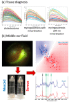
Ex vivo applications of label-free technologies in middle-ear space. (a) Differences in Raman spectra between cholesteatoma, myringosclerosis with mineralization, and myringosclerosis with no mineralization. Reprinted from [8] Discerning the differential molecular pathology of proliferative middle ear lesions using Raman spectroscopy by R. Pandey, 2015, Scientific Reports, 5(1): 13,305 (DOI: 10.1038/srep13305). CC BY 4.0. (b) Differentiating middle-ear fluids using Raman spectroscopy. Adapted from [13] Differential diagnosis of otitis media with effusion using label-free Raman spectroscopy: A pilot study by R. Pandey, 2018, J Biophotonics, 11(6): e201700259 (DOI:10.1002/jbio.201700259). Copyright 2018 by John Wiley and Sons, NJ, USA.

References

    1. Orosco R.K., Tsien R.Y., Nguyen Q.T. Fluorescence Imaging in Surgery. IEEE Rev. Biomed. Eng. 2013;6:178–187. doi: 10.1109/RBME.2013.2240294. - DOI - PMC - PubMed
    1. Singh R., Owen V., Shonde A., Kaye P., Hawkey C., Ragunath K. White light endoscopy, narrow band imaging and chromoendoscopy with magnification in diagnosing colorectal neoplasia. World J. Gastrointest. Endosc. 2009;1:45–50. doi: 10.4253/wjge.v1.i1.45. - DOI - PMC - PubMed
    1. Tan H.E.I., Maria P.L.S., Wijesinghe P., Kennedy B.F., Allardyce B.J., Eikelboom R.H., Atlas M.D., Dilley R.J. Optical Coherence Tomography of the Tympanic Membrane and Middle Ear: A Review. Otolaryngol. Neck Surg. 2018;159:424–438. doi: 10.1177/0194599818775711. - DOI - PubMed
    1. Fujimoto J.G., Pitris C., Boppart S.A., Brezinski M.E. Optical Coherence Tomography: An Emerging Technology for Biomedical Imaging and Optical Biopsy. Neoplasia. 2000;2:9–25. doi: 10.1038/sj.neo.7900071. - DOI - PMC - PubMed
    1. Zysk A.M., Nguyen F.T., Oldenburg A.L., Marks D.L., Boppart S.A. Optical coherence tomography: A review of clinical development from bench to bedside. J. Biomed. Opt. 2007;12:051403. doi: 10.1117/1.2793736. - DOI - PubMed

LinkOut - more resources