

Mitochondrial Chronic Progressive External Ophthalmoplegia
- PMID: 38391710
- PMCID: PMC10887352
- DOI: 10.3390/brainsci14020135
Mitochondrial Chronic Progressive External Ophthalmoplegia
Abstract
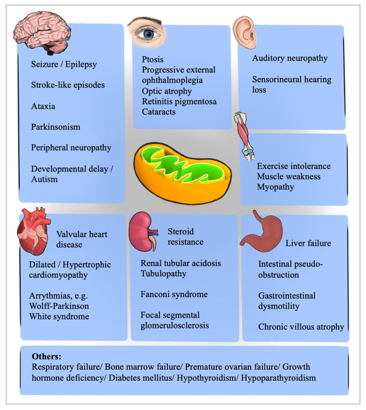
Background: Chronic progressive external ophthalmoplegia (CPEO) is a rare disorder that can be at the forefront of several mitochondrial diseases. This review overviews mitochondrial CPEO encephalomyopathies to enhance accurate recognition and diagnosis for proper management.
Methods: This study is conducted based on publications and guidelines obtained by selective review in PubMed. Randomized, double-blind, placebo-controlled trials, Cochrane reviews, and literature meta-analyses were particularly sought.
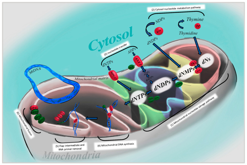
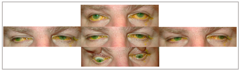
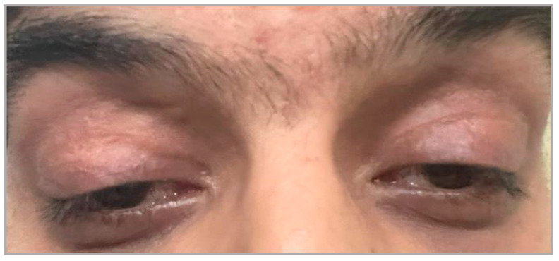
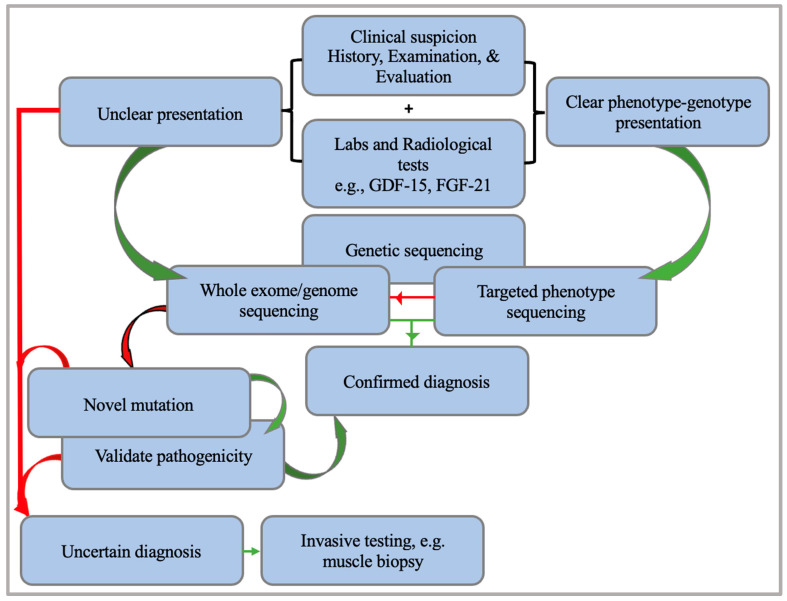
Discussion: CPEO is a common presentation of mitochondrial encephalomyopathies, which can result from alterations in mitochondrial or nuclear DNA. Genetic sequencing is the gold standard for diagnosing mitochondrial encephalomyopathies, preceded by non-invasive tests such as fibroblast growth factor-21 and growth differentiation factor-15. More invasive options include a muscle biopsy, which can be carried out after uncertain diagnostic testing. No definitive treatment option is available for mitochondrial diseases, and management is mainly focused on lifestyle risk modification and supplementation to reduce mitochondrial load and symptomatic relief, such as ptosis repair in the case of CPEO. Nevertheless, various clinical trials and endeavors are still at large for achieving beneficial therapeutic outcomes for mitochondrial encephalomyopathies.
Key messages: Understanding the varying presentations and genetic aspects of mitochondrial CPEO is crucial for accurate diagnosis and management.
Keywords: CPEO; chronic progressive external ophthalmoplegia; mitochondrial diseases.
Conflict of interest statement
The authors declare no conflicts of interest.
Figures
Similar articles
-
Mitochondrial mutations in 12S rRNA and 16S rRNA presenting as chronic progressive external ophthalmoplegia (CPEO) plus: A case report.Medicine (Baltimore). 2017 Dec;96(48):e8869. doi: 10.1097/MD.0000000000008869. Medicine (Baltimore). 2017. PMID: 29310369 Free PMC article.
-
Pontine stroke in a patient with Chronic Progressive External Ophthalmoplegia (CPEO): a case report.BMC Neurol. 2023 Jun 14;23(1):231. doi: 10.1186/s12883-023-03249-9. BMC Neurol. 2023. PMID: 37316776 Free PMC article.
-
Progressive External Ophthalmoplegia in Polish Patients-From Clinical Evaluation to Genetic Confirmation.Genes (Basel). 2020 Dec 31;12(1):54. doi: 10.3390/genes12010054. Genes (Basel). 2020. PMID: 33396418 Free PMC article.
-
Chronic progressive external ophthalmoplegia.Curr Neurol Neurosci Rep. 2002 Sep;2(5):413-7. doi: 10.1007/s11910-002-0067-5. Curr Neurol Neurosci Rep. 2002. PMID: 12169221 Review.
-
Mitochondrial encephalomyopathy with autosomal dominant inheritance: a clinical and genetic entity of mitochondrial diseases.Muscle Nerve. 1995 Jul;18(7):753-60. doi: 10.1002/mus.880180712. Muscle Nerve. 1995. PMID: 7783765 Review.
Cited by
-
Defective Neurogenesis in Lowe Syndrome is Caused by Mitochondria Loss and Cilia-related Sonic Hedgehog Defects.bioRxiv [Preprint]. 2024 Nov 1:2024.11.01.621496. doi: 10.1101/2024.11.01.621496. bioRxiv. 2024. PMID: 39553960 Free PMC article. Preprint.
-
Mapping Disorders with Neurological Features Through Mitochondrial Impairment Pathways: Insights from Genetic Evidence.Curr Issues Mol Biol. 2025 Jul 1;47(7):504. doi: 10.3390/cimb47070504. Curr Issues Mol Biol. 2025. PMID: 40728973 Free PMC article. Review.
References
-
- Yu-Wai-Man P., Clements Al Nesbitt V., Griffiths P.G., Gorman G.S., Schaefer A.M., Turnbull D.M., Taylor R.W., McFarland R. A national epidemiological study of chronic progressive external ophthalmoplegia in the United Kingdom—Molecular genetic features and neurological burden. Investig. Ophthalmol. Vis. Sci. 2014;55:5109. - PubMed
Publication types
LinkOut - more resources
Full Text Sources
