. 2024 Feb 15;13(4):344.
doi: 10.3390/cells13040344.

Morphological Changes of 3T3 Cells under Simulated Microgravity

Affiliations

Morphological Changes of 3T3 Cells under Simulated Microgravity

Minh Thi Tran et al. Cells. .

Abstract

Background: Cells are sensitive to changes in gravity, especially the cytoskeletal structures that determine cell morphology. The aim of this study was to assess the effects of simulated microgravity (SMG) on 3T3 cell morphology, as demonstrated by a characterization of the morphology of cells and nuclei, alterations of microfilaments and microtubules, and changes in cycle progression.

Methods: 3T3 cells underwent induced SMG for 72 h with Gravite®, while the control group was under 1G. Fluorescent staining was applied to estimate the morphology of cells and nuclei and the cytoskeleton distribution of 3T3 cells. Cell cycle progression was assessed by using the cell cycle app of the Cytell microscope, and Western blot was conducted to determine the expression of the major structural proteins and main cell cycle regulators.

Results: The results show that SMG led to decreased nuclear intensity, nuclear area, and nuclear shape and increased cell diameter in 3T3 cells. The 3T3 cells in the SMG group appeared to have a flat form and diminished microvillus formation, while cells in the control group displayed an apical shape and abundant microvilli. The 3T3 cells under SMG exhibited microtubule distribution surrounding the nucleus, compared to the perinuclear accumulation in control cells. Irregular forms of the contractile ring and polar spindle were observed in 3T3 cells under SMG. The changes in cytoskeleton structure were caused by alterations in the expression of major cytoskeletal proteins, including β-actin and α-tubulin 3. Moreover, SMG induced 3T3 cells into the arrest phase by reducing main cell cycle related genes, which also affected the formation of cytoskeleton structures such as microfilaments and microtubules.

Conclusions: These results reveal that SMG generated morphological changes in 3T3 cells by remodeling the cytoskeleton structure and downregulating major structural proteins and cell cycle regulators.

Keywords: 3T3 cell; cell cycle progression; cytokinesis; cytoskeleton; morphology; simulated microgravity.


Conflict of interest statement

The authors declare no conflicts of interest.

Figures

Figure 1
Figure 1
Proliferation of 3T3 cells in control and SMG groups. (A,B) Cell morphology of 3T3 cells in control and SMG groups. (C,D) FCS values for control and SMG groups (n = 5). (E) Gravite® operation in CO2 incubator. Scale bar = 223.64 µm.
Figure 2
Figure 2
Analysis of 3T3 nuclear morphology. (A) Nuclear intensity value/cell (n = 24). (B) Nuclear shape value (n = 24). (C) Nuclear area (n = 24). (D,E) Distribution of 3T3 nuclear shape values relative to nuclear intensity. (F,G) Distribution of 3T3 nuclear area values relative to nuclear intensity. Gray indicates percentage of nuclei < 2n, blue indicates percentage of nuclei in G0/G1 phase, red indicates percentage of nuclei in S phase, green indicates percentage of nuclei in G2/M phase, and yellow indicates percentage of nuclei > 4n.
Figure 3
Figure 3
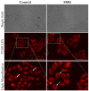
Distribution of microfilament bundles of 3T3 cells. Microfilaments were stained with phalloidin (green color), and nuclei were counterstained with H33342. White arrows indicate microvilli. Scale bar = 223.64 µm.
Figure 4
Figure 4
Distribution of microtubules of 3T3 cells. Microtubules were stained with SiR-tubulin (red color). White arrows indicate perinuclear accumulations of microtubules; dashed arrows indicate distribution of microtubules surrounding nucleus. Scale bar = 223.64 µm.
Figure 5
Figure 5
Western blot analysis of major structural proteins in 3T3 cells. α-Tubulin 3 and β-actin were downregulated in 3T3 cells under SMG (n = 3). GAPDH was used as internal control.
Figure 6
Figure 6
Cell cycle progression analysis. (A,B) Cell cycle of 3T3 cells in control and SMG groups was analyzed by cell cycle app of Cytell microscope (n = 24). Gray indicates percentage of nuclei < 2n, blue indicates percentage of nuclei in G0/G1 phase, red indicates percentage of nuclei in S phase, green indicates percentage of nuclei in G2/M phase, and yellow indicates percentage of nuclei > 4n. (C) Western blot analysis of major cell cycle-related proteins in 3T3 cells (n = 3). (D) Number of 3T3 cells was counted by cell cycle app (n = 24).
Figure 7
Figure 7
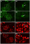
Morphology of cell division related structures in 3T3 cells: (A,A1) contractile ring in 3T3 cells in control group; (B,B1) contractile ring in 3T3 cells in SMG group; (C,C1) polar spindle in 3T3 cells in control group; (D,D1) polar spindle in 3T3 cells in SMG group. Microfilaments were counterstained using phalloidin (green), and microtubules were stained with SiR-tubulin (red). White arrows indicate contractile rings, and dashed arrows indicate polar spindles. Scale bar = 100 µm.

References

    1. Ogneva I.V. Single Cell in a Gravity Field. Life. 2022;12:1601. doi: 10.3390/life12101601. - DOI - PMC - PubMed
    1. Takahashi K., Takahashi H., Furuichi T., Toyota M., Furutani-Seiki M., Kobayashi T., Watanabe-Takano H., Shinohara M., Numaga-Tomita T., Sakaue-Sawano A., et al. Gravity sensing in plant and animal cells. NPJ Microgravity. 2021;7:2. doi: 10.1038/s41526-020-00130-8. - DOI - PMC - PubMed
    1. Conrad A.H., Stephens A.P., Conrad G.W. Effect of hexylene glycol-altered microtubule distributions on cytokinesis and polar lobe formation in fertilized eggs of Ilyanassa obsolete. J. Exp. Zool. 1994;269:188–204. doi: 10.1002/jez.1402690304. - DOI - PubMed
    1. Hung R.J., Tsao Y.D., Spauling G.F. Gravity effect on lymphocyte deformation through cell shape change. Proc. Natl. Sci. Counc. Repub. China Part B. 1995;19:19–42. - PubMed
    1. Guignandon A., Vico L., Alexandre C., Lafage-Proust M.-H. Shape Changes of Osteoblastic Cells Under Gravitational Variations during Parabolic Flight. Relationship with PGE2 Synthesis. Cell Struct. Funct. 1995;20:369–375. doi: 10.1247/csf.20.369. - DOI - PubMed

Publication types

LinkOut - more resources