

Geotemporal Fluorophore Biodistribution Mapping of Colorectal Cancer: Micro and Macroscopic Insights
- PMID: 38392057
- PMCID: PMC10887825
- DOI: 10.3390/curroncol31020063
Geotemporal Fluorophore Biodistribution Mapping of Colorectal Cancer: Micro and Macroscopic Insights
Abstract
Fluorescence-guided oncology promises to improve both the detection and treatment of malignancy. We sought to investigate the temporal distribution of indocyanine green (ICG), an exogenous fluorophore in human colorectal cancer. This analysis aims to enhance our understanding of ICG's effectiveness in current tumour detection and inform potential future diagnostic and therapeutic enhancements.
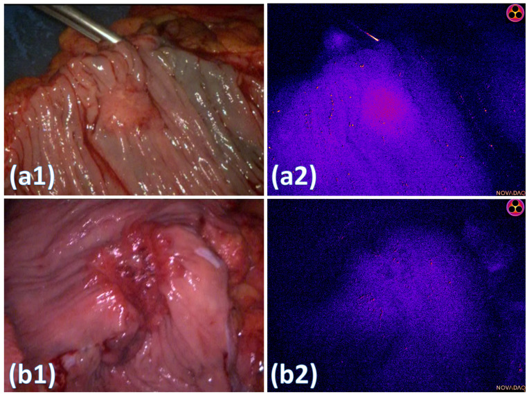
Methods: Fifty consenting patients undergoing treatment for suspected/confirmed colorectal neoplasia provided near infrared (NIR) video and imagery of transanally recorded and ex vivo resected rectal lesions following intravenous ICG administration (0.25 mg/kg), with a subgroup providing tissue samples for microscopic (including near infrared) analysis. Computer vision techniques detailed macroscopic 'early' (<15 min post ICG administration) and 'late' (>2 h) tissue fluorescence appearances from surgical imagery with digital NIR scanning (Licor, Lincoln, NE, USA) and from microscopic analysis (Nikon, Tokyo, Japan) undertaken by a consultant pathologist detailing tissue-level fluorescence distribution over the same time.
Results: Significant intra-tumoural fluorescence heterogeneity was seen 'early' in malignant versus benign lesions. In all 'early' samples, fluorescence was predominantly within the tissue stroma, with uptake within plasma cells, blood vessels and lymphatics, but not within malignant or healthy glands. At 'late' stage observation, fluorescence was visualised non-uniformly within the intracellular cytoplasm of malignant tissue but not retained in benign glands. Fluorescence also accumulated within any present peritumoural inflammatory tissue.
Conclusion: This study demonstrates the time course diffusion patterns of ICG through both benign and malignant tumours in vivo in human patients at both macroscopic and microscopic levels, demonstrating important cellular drivers and features of geolocalisation and how they differ longitudinally after exposure to ICG.
Keywords: colorectal cancer; fluorescence microscopy; fluorescence-guided surgery; fluorophore biodistribution.
Conflict of interest statement
Professor Ronan A Cahill is named on a patent filed in relation to processes for visual determination of tissue biology, receives speaker fees from Stryker Corp and Ethicon/J&J, consultancy fees from Arthrex, Diagnostic Green and Touch Surgery (Medtronic), research funding from Intuitive Corp and Medtronic as well as from the Irish Government (DTIF) in collaboration with IBM Research in Ireland, from EU Horizon 2020 in collaboration with Palliare and Horizon Europe in collaboration with Arthrex. Dr Jeffrey Dalli and Dr Niall P Hardy were employed as researchers in the D.T.I.F and Dr Jeffrey Dalli is recipient of the TESS scholarship (Malta). There are no other conflicts of interest to disclose from the remaining authors.
Figures


Similar articles
-
Digital dynamic discrimination of primary colorectal cancer using systemic indocyanine green with near-infrared endoscopy.Sci Rep. 2021 May 31;11(1):11349. doi: 10.1038/s41598-021-90089-7. Sci Rep. 2021. PMID: 34059705 Free PMC article.
-
Snapshots of lymphatic pathways in colorectal cancer surgery using near-infrared fluorescence, in vivo and ex vivo.Eur J Surg Oncol. 2021 Dec;47(12):3130-3136. doi: 10.1016/j.ejso.2021.07.025. Epub 2021 Jul 31. Eur J Surg Oncol. 2021. PMID: 34373159
-
Near-Infrared Indocyanine Green-Enhanced Fluorescence and Minimally Invasive Colorectal Surgery: Review of the Literature.Surg Technol Int. 2018 Nov 11;33:77-83. Surg Technol Int. 2018. PMID: 30029290 Review.
-
Intraoperative near infrared functional imaging of rectal cancer using artificial intelligence methods - now and near future state of the art.Eur J Nucl Med Mol Imaging. 2024 Aug;51(10):3135-3148. doi: 10.1007/s00259-024-06731-9. Epub 2024 Jun 11. Eur J Nucl Med Mol Imaging. 2024. PMID: 38858280 Free PMC article. Review.
-
The utility of indocyanine green near infrared fluorescent imaging in the identification of parathyroid glands during surgery for primary hyperparathyroidism.J Surg Oncol. 2016 Jun;113(7):771-4. doi: 10.1002/jso.24240. Epub 2016 Apr 4. J Surg Oncol. 2016. PMID: 27039880 Clinical Trial.
References
-
- Van Den Hoven P., Osterkamp J., Nerup N., Svendsen M.B.S., Vahrmeijer A., Van Der Vorst J.R., Achiam M.P. Quantitative perfusion assessment using indocyanine green during surgery—Current applications and recommendations for future use. Langenbecks Arch. Surg. 2023;408:67. doi: 10.1007/s00423-023-02780-0. - DOI - PMC - PubMed
-
- da Silva Neto E., Figueiredo P.H.M., Moro M.G., de Oliveira A.P.L., Assumpção C.B., Perina A.L.F., da Costa F.P.P., Faria E.P., de Oliveira A.C.V., Prates R.A. Use of laser-assisted indocyanine green angiography in breast reconstruction: Systematic review and meta-analysis. J. Surg. Oncol. 2020;121:759–765. doi: 10.1002/jso.25782. - DOI - PubMed
Publication types
MeSH terms
Substances
Grants and funding
LinkOut - more resources
Full Text Sources
Medical
Miscellaneous
