. 2024 Feb 14;9(2):112.
doi: 10.3390/biomimetics9020112.

Lamellar Septa-like Structured Carbonate Apatite Scaffolds with Layer-by-Layer Fracture Behavior for Bone Regeneration

Affiliations

Lamellar Septa-like Structured Carbonate Apatite Scaffolds with Layer-by-Layer Fracture Behavior for Bone Regeneration

Ahmad Nazir Taleb Alashkar et al. Biomimetics (Basel). .

Abstract

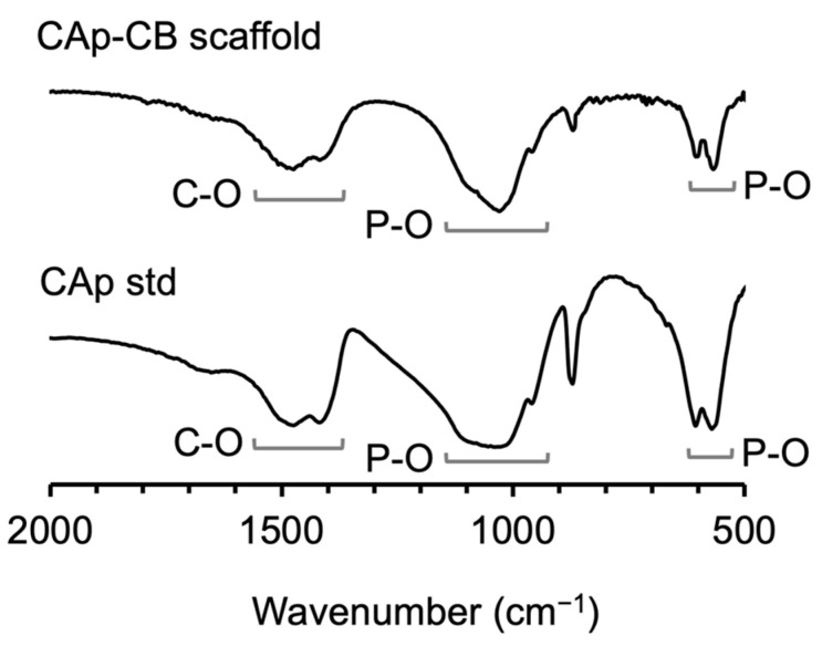
Generally, ceramics are brittle, and porosity is inversely correlated with strength, which is one of the challenges of ceramic scaffolds. Here, we demonstrate that lamellar septum-like carbonate apatite scaffolds have the potential to overcome these challenges. They were fabricated by exploiting the cellular structure of the cuttlebone, removing the organic components from the cuttlebone, and performing hydrothermal treatment. Scanning electron microscopy revealed that the scaffolds had a cellular structure with walls between lamellar septa. The interwall and interseptal sizes were 80-180 and 300-500 μm, respectively. The size of the region enclosed by the walls and septa coincided with the macropore size detected by mercury intrusion porosimetry. Although the scaffold porosity was extremely high (93.2%), the scaffold could be handled without disintegration. The compressive stress-strain curve demonstrated that the scaffolds showed layer-by-layer fracture behavior, which seemed beneficial for avoiding catastrophic failure under impact. When the scaffolds were implanted into rabbit femurs, new bone and blood vessels formed within the scaffold cells at 4 weeks. At 12 weeks, the scaffolds were almost entirely replaced with new bone. Thus, the lamellar septum-like cellular-structured carbonate apatite is a promising scaffold for achieving early bone regeneration and compression resistance.

Keywords: bioinspired materials; biomimetics; cuttlebone; scaffold; tissue engineering.


Conflict of interest statement

The authors declare no conflicts of interest.

Figures

Figure 1
Figure 1
(AC) Gross images, (DF) µ-CT images, (GL) SEM images, and (MO) high-magnification SEM images of raw CB, NaClO-treated CB, and CAp-CB scaffolds. Scale bars: (AI) 1 mm, (JL) 30 µm, and (MO) 1 µm. The green, blue, and red arrowheads indicate the walls, lamellar septa, and organic layer, respectively.
Figure 2
Figure 2
TG-DTA curves of (A) raw and (B) NaClO-treated CB cylinders. The dark and light gray arrows indicate the values of TG and DTA, respectively.
Figure 3
Figure 3
XRD patterns of the aragonite standard (std), raw and NaClO-treated CB cylinders, CAp-CB scaffold, and CAp standard (std).
Figure 4
Figure 4
FT-IR spectra of CAp-CB scaffolds and CAp std.
Figure 5
Figure 5
MIP results. (A,B) Cumulative pore volumes and (C,D) pore size distributions of raw and NaClO-treated CB cylinders and CAp-CB scaffolds.
Figure 6
Figure 6
(A) Compressive strengths of raw CB and CAp-CB (n = 8). * p < 0.005. (B) Stress–strain curves of raw CB and CAp-CB.
Figure 7
Figure 7
µ-CT images of (A) empty and (B) CAp-CB scaffold-implanted groups four weeks after surgery. (C) Empty and (D) CAp-CB scaffold-implanted groups at twelve weeks after surgery. (E) Volume percentages of new bone in the defect in the empty and CAp-CB scaffold-implanted groups at four and twelve weeks after surgery. Red lines and yellow arrowheads indicate the bone defect and new bone formed in the defect, respectively. * p < 0.005.
Figure 8
Figure 8
HE-stained sections of (A) empty and (B) CAp-CB scaffold-implanted groups four weeks post-implantation. (Upper) Images showing entire bone defects. (Lower) High-magnification images for identifying cells and tissues in the bone defects. “OBs”, “OCs”, “NB”, “BV”, “FT”, and “S” indicate osteoblasts, osteoclasts, new bone, blood vessel, fibrous tissue, and scaffold, respectively.
Figure 9
Figure 9
HE-stained sections of (A) empty and (B) CAp-CB scaffold-implanted groups twelve weeks post-implantation. (Upper) Images showing entire bone defects. (Lower) High-magnification images for identifying cells and tissues in the bone defects. “OB”, “OC”, “NB”, “BV”, “AT”, and “S” indicate osteoblast, osteoclast, new bone, blood vessel, adipose tissue, and scaffold, respectively.
Figure 10
Figure 10
Area percentages of bone newly formed in the defect area of empty and CAp-CB scaffold-implanted groups at four and twelve weeks after surgery. * p < 0.005.

References

    1. Schmidt A.H. Autologous bone graft: Is it still the gold standard? Injury. 2021;52:S18–S22. doi: 10.1016/j.injury.2021.01.043. - DOI - PubMed
    1. Gillman C.E., Jayasuriya A.C. FDA-approved bone grafts and bone graft substitute devices in bone regeneration. Mater. Sci. Eng. C. 2021;130:112466. doi: 10.1016/j.msec.2021.112466. - DOI - PMC - PubMed
    1. Govoni M., Vivarelli L., Mazzotta A., Stagni C., Maso A., Dallari D. Commercial bone grafts claimed as an alternative to autografts: Current trends for clinical applications in orthopaedics. Materials. 2021;14:3290. doi: 10.3390/ma14123290. - DOI - PMC - PubMed
    1. Janicki P., Schmidmaier G. What should be the characteristics of the ideal bone graft substitute? Combining scaffolds with growth factors and/or stem cells. Injury. 2011;42:S77–S81. doi: 10.1016/j.injury.2011.06.014. - DOI - PubMed
    1. Myeroff C., Archdeacon M. Autogenous bone graft: Donor sites and techniques. J. Bone Jt. Surg. 2011;93:2227–2236. doi: 10.2106/JBJS.J.01513. - DOI - PubMed

LinkOut - more resources