

Myocardial Late Gadolinium Enhancement (LGE) in Cardiac Magnetic Resonance Imaging (CMR)-An Important Risk Marker for Cardiac Disease
- PMID: 38392254
- PMCID: PMC10888577
- DOI: 10.3390/jcdd11020040
Myocardial Late Gadolinium Enhancement (LGE) in Cardiac Magnetic Resonance Imaging (CMR)-An Important Risk Marker for Cardiac Disease
Abstract
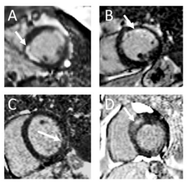
Cardiovascular magnetic resonance (CMR) has significantly revolutionized the comprehension and diagnosis of cardiac diseases, particularly through the utilization of late gadolinium enhancement (LGE) imaging for tissue characterization. LGE enables the visualization of expanded extracellular spaces in conditions such as fibrosis, fibrofatty tissue, or edema. The growing recognition of LGE's prognostic capacity underscores its importance, evident in the increasing explicit recommendations within guidelines. Notably, the contemporary characterization of cardiomyopathies relies on LGE-based scar assessment by CMR to a large extent. This review describes the pattern and prognostic value of LGE in detail for various cardiac diseases. Despite its merits, establishing LGE as a reliable risk marker encounters challenges. Limitations arise from the fact that not all diseases show LGE, and it should always be analyzed in the context of all CMR sequences and the patient's medical history. In summary, LGE stands as a robust indicator of adverse outcomes in diverse cardiovascular diseases. Its further integration into routine practice is desirable, necessitating widespread availability and application to accumulate both individual and scientific experience.
Keywords: cardiac magnetic resonance imaging; cardiomyopathy; late gadolinium enhancement; myocardial vitality; review; risk stratification.
Conflict of interest statement
The authors declare no conflicts of interest.
Figures
Similar articles
-
Incidence and prognostic significance of myocardial late gadolinium enhancement in patients with sarcoidosis without cardiac manifestation.Chest. 2014 Oct;146(4):1064-1072. doi: 10.1378/chest.14-0139. Chest. 2014. PMID: 24853830
-
[Cardiac magnetic resonance imaging and the myocardium : Differentiation between vital and nonvital tissue].Herzschrittmacherther Elektrophysiol. 2022 Sep;33(3):272-277. doi: 10.1007/s00399-022-00874-8. Epub 2022 Jul 4. Herzschrittmacherther Elektrophysiol. 2022. PMID: 35781833 Review. German.
-
Myocardial fibrosis by late gadolinium enhancement cardiovascular magnetic resonance in myotonic muscular dystrophy type 1: highly prevalent but not associated with surface conduction abnormality.J Cardiovasc Magn Reson. 2019 May 2;21(1):26. doi: 10.1186/s12968-019-0535-6. J Cardiovasc Magn Reson. 2019. PMID: 31046780 Free PMC article.
-
Evaluation of the ECG based Selvester scoring method to estimate myocardial scar burden and predict clinical outcome in patients with left bundle branch block, with comparison to late gadolinium enhancement CMR imaging.Ann Noninvasive Electrocardiol. 2017 Sep;22(5):e12440. doi: 10.1111/anec.12440. Epub 2017 Mar 1. Ann Noninvasive Electrocardiol. 2017. PMID: 28248005 Free PMC article.
-
Prognostic Value of LGE-CMR in HCM: A Meta-Analysis.JACC Cardiovasc Imaging. 2016 Dec;9(12):1392-1402. doi: 10.1016/j.jcmg.2016.02.031. Epub 2016 Jul 20. JACC Cardiovasc Imaging. 2016. PMID: 27450876 Review.
Cited by
-
Return-to-Play Post-Myocarditis for Athletes: To Play or Not to Play?Diagnostics (Basel). 2024 Oct 7;14(19):2236. doi: 10.3390/diagnostics14192236. Diagnostics (Basel). 2024. PMID: 39410640 Free PMC article. Review.
-
Cardiomyopathy in Non-Ambulatory Patients with Duchenne Muscular Dystrophy: Two Case Reports with Varying Outcomes, Considering Novel Treatments.Reports (MDPI). 2024 Dec 27;8(1):2. doi: 10.3390/reports8010002. Reports (MDPI). 2024. PMID: 40729215 Free PMC article.
-
Prevalence and Correlates of Dilated and Non-Dilated Left Ventricular Cardiomyopathy in Transfusion-Dependent Thalassemia: Data from a National, Multicenter, Observational Registry.J Cardiovasc Dev Dis. 2025 Mar 16;12(3):103. doi: 10.3390/jcdd12030103. J Cardiovasc Dev Dis. 2025. PMID: 40137101 Free PMC article.
-
Implications of Myocardial Fibrosis Burden on Left Ventricular Systolic Function in Sepsis Survivors: Insights from a Retrospective Cohort Study Using Quantitative Late Gadolinium Enhancement Cardiovascular Magnetic Resonance.J Cardiovasc Dev Dis. 2025 Aug 13;12(8):306. doi: 10.3390/jcdd12080306. J Cardiovasc Dev Dis. 2025. PMID: 40863372 Free PMC article.
-
Cardiac amyloidosis: A diagnostic challenge.Radiol Case Rep. 2024 Aug 10;19(11):4730-4735. doi: 10.1016/j.radcr.2024.07.119. eCollection 2024 Nov. Radiol Case Rep. 2024. PMID: 39228955 Free PMC article.
References
-
- Zeppenfeld K., Tfelt-Hansen J., de Riva M., Winkel B.G., Behr E.R., Blom N.A., Charron P., Corrado D., Dagres N., de Chillou C., et al. 2022 ESC Guidelines for the Management of Patients with Ventricular Arrhythmias and the Prevention of Sudden Cardiac Death: Developed by the Task Force for the Management of Patients with Ventricular Arrhythmias and the Prevention of Sudden Cardiac Death of the European Society of Cardiology (ESC) Endorsed by the Association for European Paediatric and Congenital Cardiology (AEPC) Eur. Heart J. 2022;43:3997–4126. doi: 10.1093/eurheartj/ehac262. - DOI - PubMed
Publication types
LinkOut - more resources
Full Text Sources
