

The Intrinsic Cardiac Nervous System: From Pathophysiology to Therapeutic Implications
- PMID: 38392323
- PMCID: PMC10887082
- DOI: 10.3390/biology13020105
The Intrinsic Cardiac Nervous System: From Pathophysiology to Therapeutic Implications
Abstract
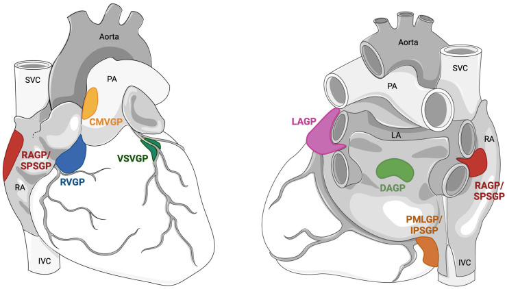
The cardiac autonomic nervous system (CANS) plays a pivotal role in cardiac homeostasis as well as in cardiac pathology. The first level of cardiac autonomic control, the intrinsic cardiac nervous system (ICNS), is located within the epicardial fat pads and is physically organized in ganglionated plexi (GPs). The ICNS system does not only contain parasympathetic cardiac efferent neurons, as long believed, but also afferent neurons and local circuit neurons. Thanks to its high degree of connectivity, combined with neuronal plasticity and memory capacity, the ICNS allows for a beat-to-beat control of all cardiac functions and responses as well as integration with extracardiac and higher centers for longer-term cardiovascular reflexes. The present review provides a detailed overview of the current knowledge of the bidirectional connection between the ICNS and the most studied cardiac pathologies/conditions (myocardial infarction, heart failure, arrhythmias and heart transplant) and the potential therapeutic implications. Indeed, GP modulation with efferent activity inhibition, differently achieved, has been studied for atrial fibrillation and functional bradyarrhythmias, while GP modulation with efferent activity stimulation has been evaluated for myocardial infarction, heart failure and ventricular arrhythmias. Electrical therapy has the unique potential to allow for both kinds of ICNS modulation while preserving the anatomical integrity of the system.
Keywords: cardiac autonomic nervous system; cardioneuroablation; ganglionated plexi ablation; intrinsic cardiac nervous system; neuromodulation.
Conflict of interest statement
The authors declare no conflicts of interest.
Figures

Similar articles
-
The role of the tripartite synapse in the heart: how glial cells may contribute to the physiology and pathophysiology of the intracardiac nervous system.Am J Physiol Cell Physiol. 2021 Jan 1;320(1):C1-C14. doi: 10.1152/ajpcell.00363.2020. Epub 2020 Oct 21. Am J Physiol Cell Physiol. 2021. PMID: 33085497 Review.
-
Cryoballoon ablation for atrial fibrillation: Effects on neuromodulation.Front Cardiovasc Med. 2022 Jul 28;9:958316. doi: 10.3389/fcvm.2022.958316. eCollection 2022. Front Cardiovasc Med. 2022. PMID: 35966567 Free PMC article. Review.
-
Ganglionated Plexi Ablation for the Treatment of Atrial Fibrillation.J Clin Med. 2020 Sep 24;9(10):3081. doi: 10.3390/jcm9103081. J Clin Med. 2020. PMID: 32987820 Free PMC article. Review.
-
Myocardial infarction induces structural and functional remodelling of the intrinsic cardiac nervous system.J Physiol. 2016 Jan 15;594(2):321-41. doi: 10.1113/JP271165. Epub 2015 Dec 15. J Physiol. 2016. PMID: 26572244 Free PMC article.
-
Evidence of structural and functional plasticity occurring within the intracardiac nervous system of spontaneously hypertensive rats.Am J Physiol Heart Circ Physiol. 2020 Jun 1;318(6):H1387-H1400. doi: 10.1152/ajpheart.00020.2020. Epub 2020 May 1. Am J Physiol Heart Circ Physiol. 2020. PMID: 32357112
Cited by
-
Cardiovascular adverse effects of immunotherapy in cancer: insights and implications.Front Oncol. 2025 Jun 18;15:1601808. doi: 10.3389/fonc.2025.1601808. eCollection 2025. Front Oncol. 2025. PMID: 40606990 Free PMC article. Review.
-
Remodeling of the Intracardiac Ganglia During the Development of Cardiovascular Autonomic Dysfunction in Type 2 Diabetes: Molecular Mechanisms and Therapeutics.Int J Mol Sci. 2024 Nov 20;25(22):12464. doi: 10.3390/ijms252212464. Int J Mol Sci. 2024. PMID: 39596529 Free PMC article. Review.
-
Antiarrhythmic Mechanisms of Epidural Blockade After Myocardial Infarction.Circ Res. 2024 Jul 19;135(3):e57-e75. doi: 10.1161/CIRCRESAHA.123.324058. Epub 2024 Jun 28. Circ Res. 2024. PMID: 38939925 Free PMC article.
-
A Comprehensive Review of a Mechanism-Based Ventricular Electrical Storm Management.J Clin Med. 2025 Jul 29;14(15):5351. doi: 10.3390/jcm14155351. J Clin Med. 2025. PMID: 40806975 Free PMC article. Review.
-
AFTER-CA: Autonomic Function Transformation and Evaluation Following Catheter Ablation in Atrial Fibrillation.J Clin Med. 2024 Sep 28;13(19):5796. doi: 10.3390/jcm13195796. J Clin Med. 2024. PMID: 39407858 Free PMC article.
References
-
- Ardell J.L., Armour J.A. Neurocardiology: Structure-Based Function. In: Terjung R., editor. Comprehensive Physiology. 1st ed. Wiley; New York, NY, USA: 2016. [(accessed on 27 December 2023)]. pp. 1635–1653. Available online: https://onlinelibrary.wiley.com/doi/10.1002/cphy.c150046. - DOI - PubMed
-
- Levy M.N. Neural control of cardiac function. Baillieres Clin. Neurol. 1997;6:227–244. - PubMed
Publication types
LinkOut - more resources
Full Text Sources
