. 2024 Feb 17;14(4):372.
doi: 10.3390/nano14040372.

In Vitro Modulation of Spontaneous Activity in Embryonic Cardiomyocytes Cultured on Poly(vinyl alcohol)/Bioglass Type 58S Electrospun Scaffolds

Affiliations

In Vitro Modulation of Spontaneous Activity in Embryonic Cardiomyocytes Cultured on Poly(vinyl alcohol)/Bioglass Type 58S Electrospun Scaffolds

Filiberto Rivera-Torres et al. Nanomaterials (Basel). .

Abstract

Because of the physiological and cardiac changes associated with cardiovascular disease, tissue engineering can potentially restore the biological functions of cardiac tissue through the fabrication of scaffolds. In the present study, hybrid nanofiber scaffolds of poly (vinyl alcohol) (PVA) and bioglass type 58S (58SiO2-33CaO-9P2O5, Bg) were fabricated, and their effect on the spontaneous activity of chick embryonic cardiomyocytes in vitro was determined. PVA/Bg nanofibers were produced by electrospinning and stabilized by chemical crosslinking with glutaraldehyde. The electrospun scaffolds were analyzed to determine their chemical structure, morphology, and thermal transitions. The crosslinked scaffolds were more stable to degradation in water. A Bg concentration of 25% in the hybrid scaffolds improved thermal stability and decreased degradation in water after PVA crosslinking. Cardiomyocytes showed increased adhesion and contractility in cells seeded on hybrid scaffolds with higher Bg concentrations. In addition, the effect of Ca2+ ions released from the bioglass on the contraction patterns of cultured cardiomyocytes was investigated. The results suggest that the scaffolds with 25% Bg led to a uniform beating frequency that resulted in synchronous contraction patterns.

Keywords: Ca2+ bioactivity; PVA/bioglass hybrid; cardiomyocytes; cell contractility patterns; electrospun scaffolds; physicochemical characterization.


Conflict of interest statement

The authors declare no conflicts of interest.

Figures

Figure 1
Figure 1
SEM micrographs of PVA scaffolds with different Bg concentrations, before and after (*) chemical crosslinking: (a,a*) 5% hybrid, (b,b*) 10% hybrid, (c,c*) 15% hybrid; (d,d*) 20% hybrid; (e,e*) 25% hybrid and (f,f*) 30% hybrid.
Figure 2
Figure 2
Shows the mean fiber diameters as a function of Bg concentration before and after chemical crosslinking with GA, measured with ImageJ software.
Figure 3
Figure 3
Elemental mapping in hybrids scaffold at 20% Bg concentration: (a) electron micrograph of PVA nanofiber, (b) Si (cyan), (c) Ca (yellow), and (d) P (green), and (e) merge image overlaying all elemental distributions on the nanofiber micrograph. Specific colors were added afterward.
Figure 4
Figure 4
FTIR spectra of PVA scaffolds with different Bg concentrations: (a) 5% hybrid; (b) 10% hybrid; (c) 15% hybrid; (d) 20% hybrid; (e) 25% hybrid; and (f) 30% hybrid.
Figure 5
Figure 5
Schematic representation of the crosslinking reaction of PVA with GA.
Figure 6
Figure 6
PVA DSC thermograms before and after crosslinking.
Figure 7
Figure 7
Fluorescence images of cardiomyocytes on hybrid PVA/Bg scaffolds, at different Bg concentrations: (ag) at rest (top row) and (a*g*) during contractile activity (bottom row). (4×) stereoscopic microscope.
Figure 8
Figure 8
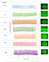
Contractile activity patterns revealed by fluorescence peaks attributable to intracellular ΔCa2+ are shown in the center panel; they were selected as regions of interest (ROIs) and are marked with a red dot on the corresponding image on the right. Right column: Fluorescence images of embryonic ventricular cardiomyocytes seeded on PVA/Bg-crosslinked scaffolds at six Bg concentrations as indicated in the left column. As can be appreciated, the different substrates do not inhibit contractile activity. However, the different Bg concentrations regulate adhesion and form separate cell aggregates (5, 10, and 15% Bg) or confluent layers (20, 25, and 30% Bg) with synchronized contractile activity in extended regions. Scale 100 μm.
Figure 9
Figure 9
(a) Diagrams of dissolution of Ca2+ ions and fiber radius versus time: dissolution of Ca2+, solid line, and fiber radius (r), dotted line. (b) Schematic of the change in fiber radius at different times.
Figure 10
Figure 10
Schematization of cardiomyocytes across the PVA/Bg scaffold surface; cardiomyocytes adhere to the surface of nanofibers in the presence of a culture medium. Released Ca2+, Si2+, and PO43− surrounding the cardiomyocyte surface and other ions from the culture medium.

References

    1. Kargozar S., Hamzehlou S., Baino F. Potential of Bioactive Glasses for Cardiac and Pulmonary Tissue Engineering. Materials. 2017;10:1429. doi: 10.3390/ma10121429. - DOI - PMC - PubMed
    1. Deaths from Cardiovascular Diseases Surges 60% Globaly on the Last 30 Years: Report. World Heart Federation; Geneva, Switzerland: 2023.
    1. Nystoriak M.A., Bhatnagar A. Cardiovascular Effects and Benefits of Exercise. Front. Cardiovasc. Med. 2018;5:135. doi: 10.3389/fcvm.2018.00135. - DOI - PMC - PubMed
    1. Freytes D.O., Santambrogio L., Vunjak-Novakovic G. Optimizing Dynamic Interactions between a Cardiac Patch and Inflammatory Host Cells. Cells Tissues Organs. 2011;195:171–182. doi: 10.1159/000331392. - DOI - PMC - PubMed
    1. Maria T.M.C., de Carvalho R.A., Sobral P.J.A., Habitante A.M.B.Q., Solorza-Feria J. The Effect of the Degree of Hydrolysis of the PVA and the Plasticizer Concentration on the Color, Opacity, and Thermal and Mechanical Properties of Films Based on PVA and Gelatin Blends. J. Food Eng. 2008;87:191–199. doi: 10.1016/j.jfoodeng.2007.11.026. - DOI

LinkOut - more resources