

Galleria mellonella Model of Coccidioidomycosis for Drug Susceptibility Tests and Virulence Factor Identification
- PMID: 38392803
- PMCID: PMC10890491
- DOI: 10.3390/jof10020131
Galleria mellonella Model of Coccidioidomycosis for Drug Susceptibility Tests and Virulence Factor Identification
Abstract
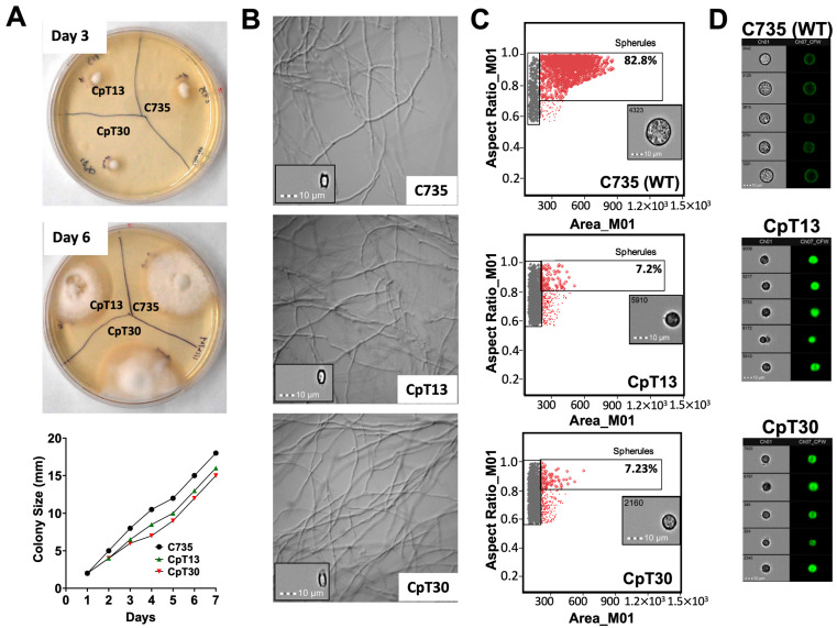
Coccidioidomycosis (CM) can manifest as respiratory and disseminated diseases that are caused by dimorphic fungal pathogens, such as Coccidioides species. The inhaled arthroconidia generated during the saprobic growth phase convert into multinucleated spherules in the lungs to complete the parasitic lifecycle. Research on coccidioidal virulence and pathogenesis primarily employs murine models typically associated with low lethal doses (LD100 < 100 spores). However, the Galleria model has recently garnered attention due to its immune system bearing both structural and functional similarities to the innate system of mammals. Our findings indicate that Coccidioides posadasii can convert and complete the parasitic cycle within the hemocoel of the Galleria larva. In Galleria, the LD100 is between 0.5 and 1.0 × 10⁶ viable spores for the clinical isolate Coccidioides posadasii C735. Furthermore, we demonstrated the suitability of this model for in vivo antifungal susceptibility tests to validate the bioreactivity of newly discovered antifungals against Coccidioides. Additionally, we utilized this larva model to screen a Coccidioides posadasii mutant library showing attenuated virulence. Similarly, the identified attenuated coccidioidal mutants displayed a loss of virulence in a commonly used murine model of coccidioidomycosis. In this study, we demonstrated that Galleria larvae can be applied as a model for studying Coccidioides infection.
Keywords: Coccidioides; Galleria mellonella; Valley Fever; drug susceptibility; fungi; mini-host; virulence factors.
Conflict of interest statement
The authors have declared that no competing interests exist.
Figures


Similar articles
-
Discovery of novel antifungal drugs via screening repurposing libraries against Coccidioides posadasii spherule initials.mBio. 2025 May 14;16(5):e0020525. doi: 10.1128/mbio.00205-25. Epub 2025 Mar 26. mBio. 2025. PMID: 40135873 Free PMC article.
-
Short-Term Memory Impairment.2024 Jun 8. In: StatPearls [Internet]. Treasure Island (FL): StatPearls Publishing; 2025 Jan–. 2024 Jun 8. In: StatPearls [Internet]. Treasure Island (FL): StatPearls Publishing; 2025 Jan–. PMID: 31424720 Free Books & Documents.
-
piv does not impact Pseudomonas aeruginosa virulence in Galleria mellonella.Microbiol Spectr. 2025 Jul;13(7):e0281124. doi: 10.1128/spectrum.02811-24. Epub 2025 May 21. Microbiol Spectr. 2025. PMID: 40396793 Free PMC article.
-
Management of urinary stones by experts in stone disease (ESD 2025).Arch Ital Urol Androl. 2025 Jun 30;97(2):14085. doi: 10.4081/aiua.2025.14085. Epub 2025 Jun 30. Arch Ital Urol Androl. 2025. PMID: 40583613 Review.
-
The Lived Experience of Autistic Adults in Employment: A Systematic Search and Synthesis.Autism Adulthood. 2024 Dec 2;6(4):495-509. doi: 10.1089/aut.2022.0114. eCollection 2024 Dec. Autism Adulthood. 2024. PMID: 40018061 Review.
Cited by
-
Comparison of Morphological, Virulence, and Antifungal Susceptibility Characteristics Within Aspergillus Lentulus.Infect Drug Resist. 2025 Jul 30;18:3761-3769. doi: 10.2147/IDR.S532973. eCollection 2025. Infect Drug Resist. 2025. PMID: 40756688 Free PMC article.
-
Inferring the composition of a mixed culture of natural microbial isolates by deep sequencing.bioRxiv [Preprint]. 2024 Aug 5:2024.08.05.606565. doi: 10.1101/2024.08.05.606565. bioRxiv. 2024. PMID: 39149389 Free PMC article. Preprint.
-
Virulence and biological characteristics of Talaromyces wortmannii isolated from deep-seated dermatomycosis by in vitro and in vivo evaluation.BMC Infect Dis. 2025 Jul 1;25(1):884. doi: 10.1186/s12879-025-11261-2. BMC Infect Dis. 2025. PMID: 40596872 Free PMC article.
-
Galleria mellonella as an Invertebrate Model for Studying Fungal Infections.J Fungi (Basel). 2025 Feb 18;11(2):157. doi: 10.3390/jof11020157. J Fungi (Basel). 2025. PMID: 39997451 Free PMC article. Review.
-
"Select and Resequence" Methods Enable a Genome-Wide Association Study of the Dimorphic Human Fungal Pathogen Coccidioides posadasii.Genome Biol Evol. 2025 Jul 3;17(7):evaf135. doi: 10.1093/gbe/evaf135. Genome Biol Evol. 2025. PMID: 40611625 Free PMC article.
References
-
- Engelthaler D.M., Roe C.C., Hepp C.M., Teixeira M., Driebe E.M., Schupp J.M., Gade L., Waddell V., Komatsu K., Arathoon E., et al. Local Population Structure and Patterns of Western Hemisphere Dispersal for Coccidioides spp., the Fungal Cause of Valley Fever. mBio. 2016;7:e00550-16. doi: 10.1128/mBio.00550-16. - DOI - PMC - PubMed
-
- Litvintseva A.P., Marsden-Haug N., Hurst S., Hill H., Gade L., Driebe E.M., Ralston C., Roe C., Barker B.M., Goldoft M., et al. Valley fever: Finding new places for an old disease: Coccidioides immitis found in Washington State soil associated with recent human infection. Clin. Infect. Dis. 2015;60:e1–e3. doi: 10.1093/cid/ciu681. - DOI - PMC - PubMed
-
- Chiller T. Vaccine Strategies for Endemic Fungal Pathogens. NIAID; Rockville, MD, USA: 2019. Overview of endemic mycosis.
-
- Pappagianis D. Clinical presentation of infectious entities. In: Einstein H., editor. Coccidioidomycosis. National Foundation for Infectious Disease; Washington, DC, USA: 1996. pp. 9–11.
Grants and funding
LinkOut - more resources
Full Text Sources
Research Materials
