. 2024 Feb 16;14(2):126.
doi: 10.3390/metabo14020126.

Circadian Disruption across Lifespan Impairs Glucose Homeostasis and Insulin Sensitivity in Adult Mice

Affiliations

Circadian Disruption across Lifespan Impairs Glucose Homeostasis and Insulin Sensitivity in Adult Mice

Tracy K Her et al. Metabolites. .

Abstract

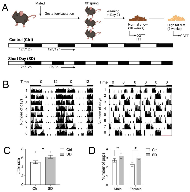
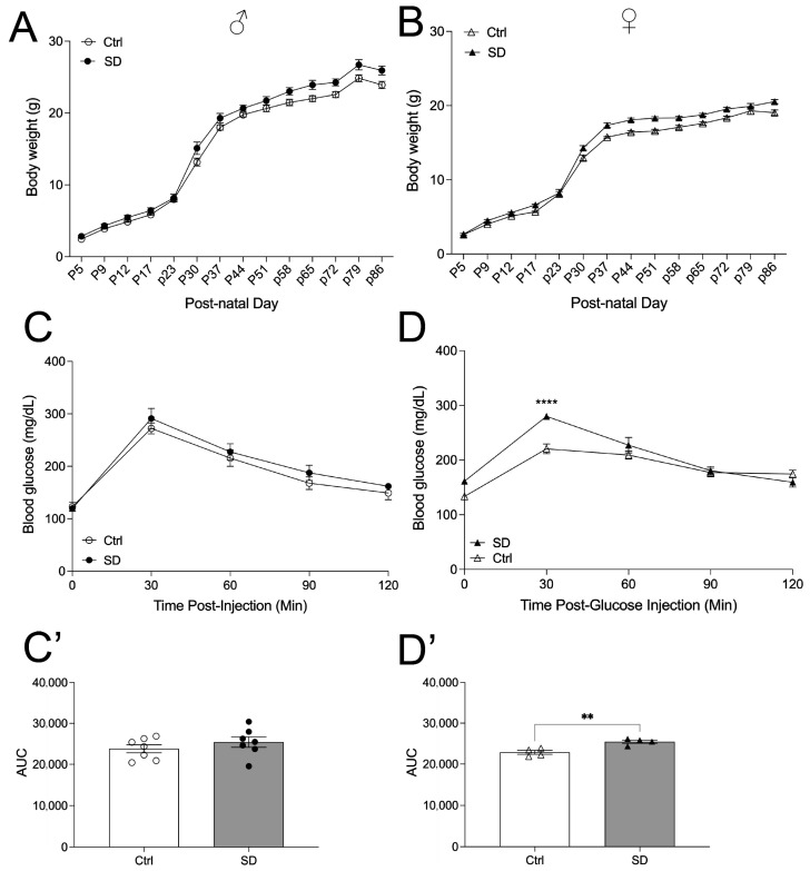
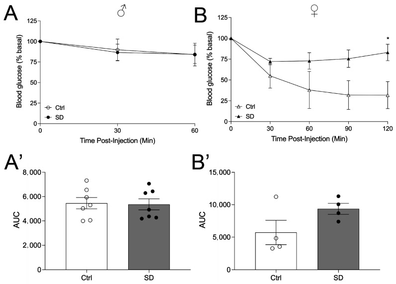
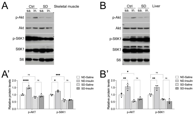
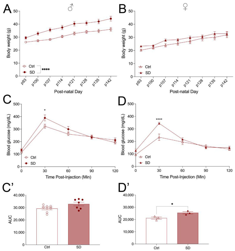
Circadian rhythm disruption is associated with impaired glucose homeostasis and type 2 diabetes. For example, night shift work is associated with an increased risk of gestational diabetes. However, the effects of chronic circadian disruption since early life on adult metabolic health trajectory remain unknown. Here, using the "Short Day" (SD) mouse model, in which an 8 h/8 h light/dark (LD) cycle was used to disrupt mouse circadian rhythms across the lifespan, we investigated glucose homeostasis in adult mice. Adult SD mice were fully entrained into the 8 h/8 h LD cycle, and control mice were entrained into the 12 h/12 h LD cycle. Under a normal chow diet, female and male SD mice displayed a normal body weight trajectory. However, female but not male SD mice under a normal chow diet displayed glucose intolerance and insulin resistance, which are associated with impaired insulin signaling/AKT in the skeletal muscle and liver. Under high-fat diet (HFD) challenges, male but not female SD mice demonstrated increased body weight gain compared to controls. Both male and female SD mice developed glucose intolerance under HFD. Taken together, these results demonstrate that environmental disruption of circadian rhythms contributes to obesity in a sexually dimorphic manner but increases the risk of glucose intolerance and insulin resistance in both males and females.

Keywords: circadian disruption; glucose; insulin; mice; obesity.


Conflict of interest statement

The authors declare no conflicts of interest.

Figures

Figure 1
Figure 1
Experimental design. (A) Schematic timeline of the experiments in the current study. (B) Actograms of wheel-running activities of C57BL/6J mice under the 12 h/12 h (control, Ctrl and 8 h/8 h light/dark (short day, SD) cycles. (C) Litter size from Ctrl and SD dams. (D) Number of males and female pups born to Ctrl and SD dams. Statistical analysis was performed using an unpaired, two-tailed, Student’s t-test. * p ≤ 0.05, ns, not significant.
Figure 2
Figure 2
Female SD mice demonstrate impaired glucose tolerance on a normal chow diet (NCD). Male (circles) body weight (A) and female (triangles) body weight (B) of Ctrl and SD mice up to 86 days of postnatal age (males n = 7 and females n = 5–7). Oral glucose tolerance test (OGTT; 2 g/kg glucose) of males ((C), n = 6–7) and females ((D), n = 4) performed at 7–10 weeks of age. Area under curve analysis of OGTT males (C’) and females (D’). Statistical analysis performed using an unpaired, two-tailed, Student’s t-test or a 2-way ANOVA. ** p ≤ 0.01, **** p ≤ 0.0001.
Figure 3
Figure 3
Female SD mice show impaired insulin sensitivity. The intraperitoneal insulin tolerance test (ITT; 1.00 U/kg insulin, i.p.) was performed at 7–10 weeks of age in males (circles) ((A), n = 7) and females (triangles) ((B), n = 4). AUC analysis of ITT in males (A’) and females (B’). Statistical analysis performed using an unpaired, two-tailed, Student’s t-test, or 2-way ANOVA. * p ≤ 0.05.
Figure 4
Figure 4
Female SD mice show a decreased insulin signaling response in the skeletal muscle and liver. (A) Representative image of Western blots of p-AKT, AKT, p-S6K1, and S6K1 of skeletal muscle lysates after stimulation with saline (sa) or insulin (in) with quantification of p-AKT and p-S6K1 in (A’). (B) Representative image of Western blots of p-AKT, AKT, p-S6K1, and S6K1 of liver lysates after stimulation with sa or in with quantification of p-AKT and p-S6K1 in (B’). Skeletal muscle and liver tissue were harvested from 8-week-old female SD mice 5 min after administration of 1.0 U/kg insulin i.p. The levels of p-AKT and p-S6K1 were normalized based on the levels of total AKT and S6K1. Ribosomal proteins S6 (S6) and β-actin were used as loading controls. Statistical analysis was performed using a 2-way ANOVA. * p ≤ 0.05, ** p ≤ 0.01, *** p ≤ 0.001, **** p ≤ 0.0001, ns, not significant.
Figure 5
Figure 5
Both male and female SD mice demonstrate glucose intolerance on a high-fat diet. Male (circles) body weight (A) and female (triangles) body weight (B) of Ctrl and SD mice (males n = 7, females n = 7). Oral glucose tolerance test (OGTT; 2 g/kg glucose) of males ((C), n = 7) and females ((D), n = 3) performed at 14–16 weeks of age. AUC analysis of OGTT males (C’) and females (D’). Statistical analysis was performed using an unpaired, two-tailed, Student’s t-test or 2-way ANOVA. * p ≤ 0.05, **** p ≤ 0.0001.

References

    1. Reppert S.M., Weaver D.R. Coordination of circadian timing in mammals. Nature. 2002;418:935–941. doi: 10.1038/nature00965. - DOI - PubMed
    1. Rosbash M., Bradley S., Kadener S., Li Y., Luo W., Menet J.S., Nagoshi E., Palm K., Schoer R., Shang Y., et al. Transcriptional feedback and definition of the circadian pacemaker in Drosophila and animals. Cold Spring Harb. Symp. Quant. Biol. 2007;72:75–83. doi: 10.1101/sqb.2007.72.062. - DOI - PubMed
    1. Costello H.M., Gumz M.L. Circadian Rhythm, Clock Genes, and Hypertension: Recent Advances in Hypertension. Hypertension. 2021;78:1185–1196. doi: 10.1161/HYPERTENSIONAHA.121.14519. - DOI - PMC - PubMed
    1. Silva B.S.A., Uzeloto J.S., Lira F.S., Pereira T., Coelho E.S.M.J., Caseiro A. Exercise as a Peripheral Circadian Clock Resynchronizer in Vascular and Skeletal Muscle Aging. Int. J. Environ. Res. Public Health. 2021;18:12949. doi: 10.3390/ijerph182412949. - DOI - PMC - PubMed
    1. Mohawk J.A., Green C.B., Takahashi J.S. Central and peripheral circadian clocks in mammals. Annu. Rev. Neurosci. 2012;35:445–462. doi: 10.1146/annurev-neuro-060909-153128. - DOI - PMC - PubMed