. 2024 Feb 23;10(8):eadj1640.
doi: 10.1126/sciadv.adj1640. Epub 2024 Feb 23.

The incredible bulk: Human cytomegalovirus tegument architectures uncovered by AI-empowered cryo-EM

Affiliations

The incredible bulk: Human cytomegalovirus tegument architectures uncovered by AI-empowered cryo-EM

Jonathan Jih et al. Sci Adv. .

Abstract

The compartmentalization of eukaryotic cells presents considerable challenges to the herpesvirus life cycle. The herpesvirus tegument, a bulky proteinaceous aggregate sandwiched between herpesviruses' capsid and envelope, is uniquely evolved to address these challenges, yet tegument structure and organization remain poorly characterized. We use deep-learning-enhanced cryogenic electron microscopy to investigate the tegument of human cytomegalovirus virions and noninfectious enveloped particles (NIEPs; a genome packaging-aborted state), revealing a portal-biased tegumentation scheme. We resolve atomic structures of portal vertex-associated tegument (PVAT) and identify multiple configurations of PVAT arising from layered reorganization of pUL77, pUL48 (large tegument protein), and pUL47 (inner tegument protein) assemblies. Analyses show that pUL77 seals the last-packaged viral genome end through electrostatic interactions, pUL77 and pUL48 harbor a head-linker-capsid-binding motif conducive to PVAT reconfiguration, and pUL47/48 dimers form 45-nm-long filaments extending from the portal vertex. These results provide a structural framework for understanding how herpesvirus tegument facilitates and evolves during processes spanning viral genome packaging to delivery.


Figures

Fig. 1.
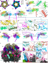
Fig. 1.. IsoNet-enhanced cryo-ET shows portal-referenced tegument in HCMV enveloped particles.
(A) 2D slices of a reconstructed tomogram with missing-wedge correction by IsoNet (49) show HCMV virions and NIEPs with asymmetrically distributed tegument compartments (green arrows). Some NIEP capsids contain a spherical scaffold core, whereas others contain scattered density likely arising from broken scaffold proteins and/or incompletely packaged dsDNA genome. Portal dodecamer (purple arrow) is clearly visible in a NIEP. (B) NIEP boxed in (A) was computationally extracted and segmented. (C) Capsomer features are visible underneath the envelope. (D) Clipped view of (C) shows portal dodecamer and PVAT. (E) Mask diameter, d, is used to approximate the capsid’s surface area (SA). Coordinates of the portal, capsid center, and bulky tegument centroid are recorded using magenta, cyan, and lime green 3D markers, respectively. (F) Capsid polar angles are defined with respect to the portal vertex, taken to be the “north pole,” or 0°. The portal-opposite vertex, or the capsid’s “south pole” is 180°. (G) The PCT angle is defined as the angle between two vectors connecting capsid center to portal (cyan to magenta), and capsid center to the bulky tegument centroid (cyan to lime green). (H) Histogram showing measured PCT angular frequency sampled from 49 HCMV particles where portal complexes could be reasonably identified. (I) Histogram showing PCT angular distribution normalized to capsid surface area, revealing a tendency of tegument to localize above the portal vertex.
Fig. 2.
Fig. 2.. Cryo-EM reveals fully-tegumented PVAT structure in virions and NIEPs.
(A and B) Global portal vertex-resolved reconstructions of VC1 virion (A) and NC1 NIEP (B) capsid. (C) Tropic penton vertices show preferred tegumentation at portal-proximal CVSC-binding-registers (black arrows). Dashed line is an equatorial drawn for reference. (D to G) Subparticle reconstructions of VC1 PVAT [(D) and (E)] and NC1 PVAT [(F) and (G)] segmented and colored as in (H). Contours chosen to best display overall structure features. (H) Exploded view showing components and layered organization of VC1-decorated portal vertex. AU1 to AU5 denotes asymmetric units 1 to 5.
Fig. 3.
Fig. 3.. Electrostatic interactions characterize structure and function of pUL77 portal cap.
(A) Overview of VC1 portal vertex shows two-layered pUL77 portal cap docked above pUL93 and portal channel. Models shown in cylinder-stub [portal model PDB 7ETM (46)]. (B) Atomic models of pUL77-u/l and local density fit. Rainbow coloring N to C (red to blue). Dashed circles denote termini. (C) Top view of portal cap, colored to demarcate asymmetric unit (AU) contributions. Five pUL77-u copies comprise the upper pentamer. (D) Adjacent pUL77-u pentamer subunits interlock via a positively charged “groove” (blue surface) and negatively charged “ridge” (red surface). (E) Ridge-forming residues of AU1 pUL77-u shown in lime green. (F) Groove-forming residues of AU2 pUL77-u shown in yellow. (G) Interfacing residue stretches between pUL77-l and pUL93 colored in maroon and dark green. (H) Interacting residues between AU2 pUL77-u and AU1 pUL77-l denoted by colored dots. (I) Clipped view of pUL77 portal cap rendered as electrostatic surface reveals a positively charged inverted “funnel” that retains DNA terminus’s rod-like density, shown fitted with ideal B-form DNA. (J) Top/bottom views of portal cap show the DNA-interfacing funnel is more positively charged (bottom view). (K) Lysine and arginine residues from pUL77-u/l line the funnel. Dashed circles denote exposed pUL77 C termini. aa., amino acids.
Fig. 4.
Fig. 4.. pUL47/48 dimerization underlies architecture of VC1/NC1 large tegument layers.
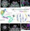
(A) Top/bottom views of VC1 LTLs. (B and C) Atomic models of pUL47-l (B) and pUL48-l (C) and respective local density fit. Rainbow coloring N to C (red to blue). Dashed circles denote termini. (D) Cylinder-stub models of pUL47/48-l dimer colored by domain. Dashed lines demarcate dimer interface. pUL47 domains: ApD, apical domain; SpD, spine domain; LTBD, large-tegument–binding domain; LSD, linker-stabilizing domain. pUL48 domains: coiled-coil motif; CDD, capsid-distal domain; CPD, capsid-proximal domain; linker–CBD, linker–capsid-binding domain. (E) Primary subunit interface of pUL47/48 dimer. Interacting residues denoted by colored dots. (F) The linker of pUL48-l’s linker–CBD (magenta) forms an additional interface with pUL47-l and is fixed to CDD via disulfide bridge (C2179-C1633, highlighted yellow). (G) Putative kinesin-binding motif WD4 in α-herpesvirus large tegument protein (24) maps to HCMV pUL48 residues TQWPAM (purple), which reside on an exposed loop of pUL48’s CPD β barrel. (H) In each AU, an upper and lower pUL47/48 dimer stack to form a tetramer. TQWPAM is exposed on the upper pUL47/48 dimer. (I) Extensive interactions between pUL48-u and pUL48-l facilitate tetramer assembly. Left: pUL48-u shown as violet silhouette; interacting residues of pUL48-l shown as pink spheres. Right: pUL48-l shown as blue silhouette; interacting residues of pUL48-u shown as purple spheres. (J) Full atomic structure of VC1 PVAT and surrounding capsid. PVAT AU1 depicted in cylinder-stub. (K and L) pUL47-l drives inter-AU assembly through interactions with an adjacent AU’s pUL47-u (K) and pUL48-l (L). (M and N) SCP-VC1 PVAT interactions occur at each AU’s CBD helix bundle (M) and pUL48-l CPD (N). aa., amino acids.
Fig. 5.
Fig. 5.. Structures of partially tegumented portal vertices demonstrate PVAT plasticity.
(A and B) Global portal vertex-resolved reconstructions of VC2 virion (A) and NC2-inv NIEP (B) capsid. (C to F) Subparticle reconstructions of VC2 PVAT [(C) and (D)] and NC2-inv PVAT [(E) and (F)] segmented and colored as in (G). Contours chosen to best display overall structure features. (G) Exploded view showing components and layered organization of NC2-inv–decorated portal vertex. Low occupancy subunits displayed as models docked in mesh density. (H) NC2-inv pUL48-l interactions with pUL77 and low-occupancy pUL47-l (docked in mesh density), with pUL48-l and pUL47-l colored by domain. pUL47-l’s relative orientation to pUL48-l is unchanged from that observed in VC1/NC1 pUL47/48 dimers. (I and J) pUL48-l interactions with pUL77, with interfacing residue stretches in pUL77 colored purple, and interacting residues in pUL48-l denoted by colored dots. (K) Full atomic structure of NC2-inv pUL48/77 decamer and surrounding capsid (low-occupancy components of NC2-inv PVAT removed for clarity). PVAT AU1 depicted in cylinder-stub. pUL77 pentamer depicted as electrostatic surface, showing positively charged (blue), normally DNA-interacting residues facing outward, true to the inverted name. (L) NC2-inv pUL48/77 decamer, flipped 180° and rigid-body fit into VC2 PVAT density. (M) Inter-AU interactions in pUL48/77 decamer between adjacent pUL48-l copies. (N) pUL48-l (interacting residues yellow) contacts neighboring P1 MCP tower (interacting residues light gray). (O) pUL48-l and its low occupancy copy of pUL47-l (yellow mesh) fit into a valley-like cleft, likely increasing the stability of pUL48/77 decamer in NC2-inv. aa., amino acids.
Fig. 6.
Fig. 6.. pUL47/pUL48 coiled-coil repeats manifest as filamentous PVAT densities.
(A and B) Top (A) and side (B) views of VC1 PVAT with docked composite [i.e., AlphaFold2 (61) and cryo-EM] models. Ten sets of filaments arising from 10 pUL47/48 dimers decorate the portal vertex. Atomic models of surrounding SCP [PDB 5VKU (38)] and pp150 with bound tRNA [PDB 7LJ3 (45)] are shown to contextualize the filament environment. (C) Unassigned fibrillar density [cyan arrows; also in (A) and (B)] connects pUL48-u’s TQWPAM motif to its own CCR filament hairpin at low contours. (D) Composite pUL47/48 structure of cryo-EM atomic models and AlphaFold2-predicted models. (E) Schematic depicting domain organization and modeling of full-length pUL47 and pUL48. Colors correspond to (D). (F) AlphaFold2-predicted models of pUL47 C-terminal and pUL48 N-terminal fragments used for density-guided fitting. Models colored by predicted local-distance difference test (plDDT) confidence scores from low (red) to high (blue). (G and H) Top (G) and side (H) views of NC2-inv PVAT with docked composite models, as in (A) and (B). (I) NC2-inv pUL48-l harbors truncated CCR filamentous density. Unmodeled density adjacent to the truncated filament belongs to a pp150’s disordered C terminus. Mesh density in (A) to (C) and (G) to (I) is of VC1 and NC2-inv global capsid reconstructions, respectively. aa., amino acids.
Fig. 7.
Fig. 7.. Structure of the penton vertex CVSC-binding-register.
(A) Workflow for generating the C1 reconstruction of a penton vertex with one CVSC-bound CVSC-binding-register. (B) Comparison of Ta triplex underneath penton vertex CVSC (left) and portal vertex CVSC (right) reveals differences in density in penton-proximal Tri1’s trunk domain (black arrow). Both Ta triplexes are rotated ~120° counterclockwise relative to non–CVSC-bound Ta triplexes. (C) Filtering the CVSC-bound penton vertex map permits a poly-alanine backbone trace of penton-proximal CVSC-bound Ta Tri1’s trunk domain (~amino acids 40 to 160), which when superposed with portal-proximal Ta Tri1, highlights conformational rearrangement of penton-proximal CVSC-bound Ta Tri1’s trunk domain. (D and E) Globular head-like tegument densities extending from CVSC helices are visible above the CVSC-binding-register in filtered CVSC-bound penton vertex maps at low contours. These fit two docked copies of pUL47/48 dimer. (F) The more penton-distal pUL47/48 dimer has noticeably weaker density. (G and H) Docking models of penton CVSC with its associated pUL47/48 dimers into the northern (G) and southern (H) tropic penton vertices show that pUL48’s globular head and pUL47 account for the preferred tegumentation of portal-proximal CVSC-binding-registers at penton vertices. Dashed lines represent equatorials delineating portal-proximal and portal-distal sides of each penton vertex.
Fig. 8.
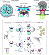
Fig. 8.. A highly configurable tegument may facilitate capsid traversal through cellular architecture.
(A) Density map is a subtomogram reconstruction (EMD-22207) from an in vitro large unilamellar vesicle (LUV) system previously used to study herpesvirus primary envelopment during nuclear egress. Recombinantly expressed HSV-1 pUL25 was found to form star-shaped clusters of density when introduced to an HSV-1 NEC lattice seeded on the LUV surface (16). Capsid-bound pUL25 was proposed to initiate NEC-mediated budding. Our capsid-derived HCMV pUL77 pentamer model shows good fit with star-shaped HSV-1 pUL25 density. Rotated side view shows a cartoon illustrating how linker–CBDs might allow pUL48 heads to flexibly fold aside, thus permitting pUL77 pentamer-NEC interactions required for primary envelopment. (B) Flowchart illustrating the proposed evolution of PVAT structure during virus maturation. Our data show enveloped virus particles contain a heterogeneous population of PVAT states, which analyses suggest may be an equilibrium of reconfiguring states, as indicated by blue double-headed arrows. Break bars in arrows represent processes not illustrated and for which PVAT structure is unknown and cannot be inferred. Nuclear capsids as illustrated here reflect the current understanding of nascent virion and NIEP formation, including the attachment of CVSC components to nuclear capsids, and NEC-mediated primary envelopment. (C) Diagram illustrating how portal-biased tegumentation may facilitate proper portal vertex orientation during NPC docking via tegument-nucleoporin interactions.

References

    1. Lazzarotto T., Guerra B., Gabrielli L., Lanari M., Landini M. P., Update on the prevention, diagnosis and management of cytomegalovirus infection during pregnancy. Clin. Microbiol. Infect. 17, 1285–1293 (2011). - PubMed
    1. U. S. Akpan, L. S. Pillarisetty, in StatPearls. (Treasure Island, 2022).
    1. W. Britt, in Human Cytomegalovirus, T. E. Shenk, M. F. Stinski, Eds. (Springer Berlin Heidelberg, 2008), pp. 417–470.
    1. Griffiths P., Reeves M., Pathogenesis of human cytomegalovirus in the immunocompromised host. Nat. Rev. Microbiol. 19, 759–773 (2021). - PMC - PubMed
    1. Wang H., Peng G., Bai J., He B., Huang K., Hu X., Liu D., Cytomegalovirus infection and relative risk of cardiovascular disease (ischemic heart disease, stroke, and cardiovascular death): A meta-analysis of prospective ptudies up to 2016. J. Am. Heart Assoc. 6, e005025 (2017). - PMC - PubMed

Publication types

Substances