

Teres minor denervation and pathologies resulting in shoulder joint instability and rotator cuff tears: A retrospective cross-sectional MRI study
- PMID: 38394498
- PMCID: PMC11309639
- DOI: 10.1097/MD.0000000000037232
Teres minor denervation and pathologies resulting in shoulder joint instability and rotator cuff tears: A retrospective cross-sectional MRI study
Abstract
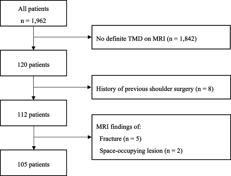
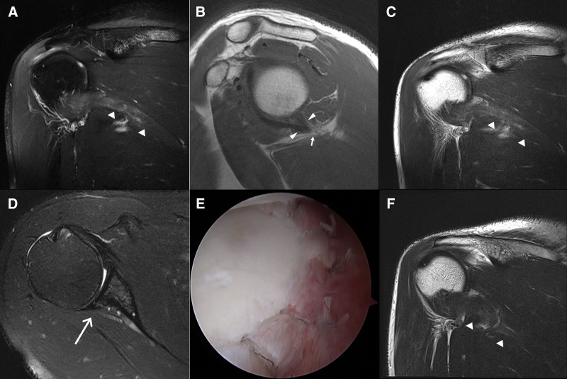
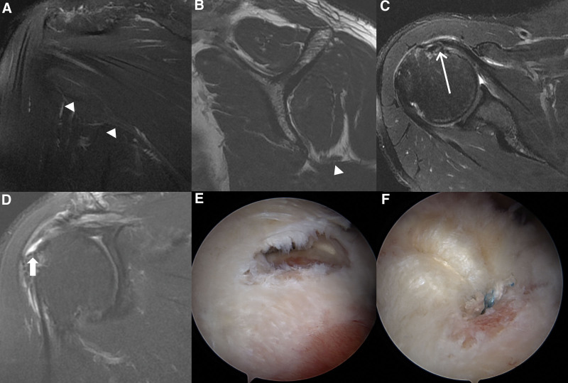
Teres minor denervation (TMD) has gained increasing attention in recent years, particularly with the advent of magnetic resonance imaging (MRI). The potential association between TMD and shoulder instability or rotator cuff tear remains a subject of interest in the orthopedic community. In this retrospective and cross-sectional study, authors aim to investigate the potential association between TMD and shoulder instability or rotator cuff tears. Authors retrospectively analyzed MRI findings from 105 patients with TMD, focusing on rotator cuff pathologies, posterior labrocapsular complex (PLCC) tears, and posteroinferior glenohumeral joint capsule alterations. Authors assessed the association between TMD and rotator cuff and PLCC tears. For the multivariate analysis, partial proportional odds models were constructed for subscapularis (SSC) and SSP tears. Rotator cuff tears were present in 82.9% of subjects, with subscapularis (SSC) tears being the most frequent (77.1%). A significant association was observed between TMD and rotator cuff pathology (P = .002). PLCC tears were found in 82.3% of patients, and humeral position relative to the osseous glenoid was noted in 60% of patients with TMD. A significant association was identified between TMD and shoulder instability or labral/capsular abnormalities (P < .001). More than half of the cases exhibited a long tethering appearance toward the axillary neurovascular bundle on T1-weighted sagittal images. Our findings suggest that TMD is significantly associated with rotator cuff tears and shoulder instability. This study highlights the importance of identifying and treating PLCC tears in patients with TMD to address shoulder instability. Further research is needed to elucidate the role of TMD in the pathogenesis of shoulder instability and rotator cuff pathology.
Copyright © 2024 the Author(s). Published by Wolters Kluwer Health, Inc.
Conflict of interest statement
The authors have no funding and conflicts of interest to disclose.
Figures

Similar articles
-
Rotator cuff muscle imbalance associates with shoulder instability direction.J Shoulder Elbow Surg. 2023 Jan;32(1):33-40. doi: 10.1016/j.jse.2022.06.022. Epub 2022 Aug 9. J Shoulder Elbow Surg. 2023. PMID: 35961497
-
Analysis of glenohumeral morphological factors for anterior shoulder instability and rotator cuff tear by magnetic resonance imaging.J Orthop Surg (Hong Kong). 2018 May-Aug;26(2):2309499018768100. doi: 10.1177/2309499018768100. J Orthop Surg (Hong Kong). 2018. PMID: 29635957
-
Anterior Shoulder Instability in the Aging Population: MRI Injury Pattern and Management.AJR Am J Roentgenol. 2021 May;216(5):1300-1307. doi: 10.2214/AJR.20.24011. Epub 2021 Feb 24. AJR Am J Roentgenol. 2021. PMID: 32783552
-
Reverse shoulder arthroplasty for primary glenohumeral osteoarthritis: significantly different characteristics and outcomes in shoulders with intact vs. torn rotator cuff.J Shoulder Elbow Surg. 2024 Apr;33(4):850-862. doi: 10.1016/j.jse.2023.07.027. Epub 2023 Aug 25. J Shoulder Elbow Surg. 2024. PMID: 37633591 Review.
-
[Shoulder instability and rotator cuff tear].Orthopade. 2009 Jan;38(1):70-4. doi: 10.1007/s00132-008-1356-8. Orthopade. 2009. PMID: 19139844 Review. German.
References
-
- Williams MD, Edwards TB, Walch G. Understanding the importance of the teres minor for shoulder function: functional anatomy and pathology. J Am Acad Orthop Surg. 2018;26:150–61. - PubMed
-
- Melis B, DeFranco MJ, Lädermann A, et al. . The teres minor muscle in rotator cuff tendon tears. Skeletal Radiol. 2011;40:1335–44. - PubMed
-
- Cahill BR, Palmer RE. Quadrilateral space syndrome. J Hand Surg A. 1983;8:65–9. - PubMed
-
- Fuchs B, Weishaupt D, Zanetti M, et al. . Fatty degeneration of the muscles of the rotator cuff: assessment by computed tomography versus magnetic resonance imaging. J Shoulder Elbow Surg. 1999;8:599–605. - PubMed
-
- Sofka CM, Lin J, Feinberg J, et al. . Teres minor denervation on routine magnetic resonance imaging of the shoulder. Skeletal Radiol. 2004;33:514–8. - PubMed
MeSH terms
LinkOut - more resources
Full Text Sources
Medical
