

Ultrasound for monitoring different stages of post-transplant lymphoproliferative disorder in a transplanted kidney: A case report and review of the literature
- PMID: 38394510
- PMCID: PMC11309683
- DOI: 10.1097/MD.0000000000036206
Ultrasound for monitoring different stages of post-transplant lymphoproliferative disorder in a transplanted kidney: A case report and review of the literature
Abstract
Rationale: Post-transplant lymphoproliferative disorder (PTLD) is a well-recognized, but uncommon complication in patients with kidney transplantation, which poses challenges in diagnosis and poor prognosis due to its low incidence and nonspecific clinical manifestations. As a routine follow-up examination method for kidney transplant patients, ultrasound (US) plays a significant role in the diagnosis of PTLD. Therefore, it is critical to evaluate the ultrasonic characteristics of PTLD in transplanted kidney patients for early detection and diagnosis.
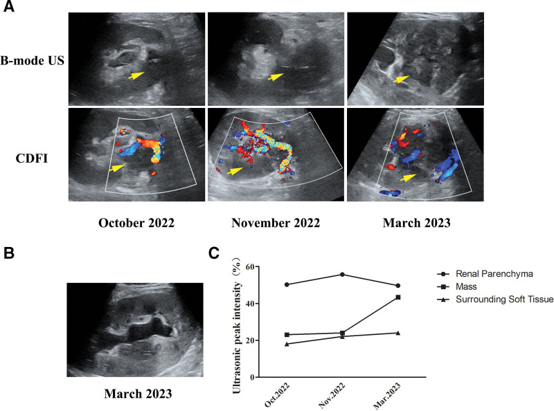
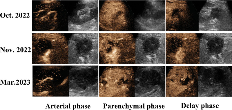
Patient concerns: A 59-year-old female patient was unexpectedly found with a mass in the hilum of the transplanted kidney 12th month after transplantation, which gradually grew up in the following 4 months. The latest US examination found hydronephrosis. Contrast-enhanced ultrasound (CEUS) demonstrated a hypo-enhancement pattern in arterial and parenchymal phases and showed a new irregular area lacking perceivable intensification within the mass, which was considered necrosis. Meanwhile, the patient developed an acute increase in serum creatinine from 122 to 195 μmol/L.
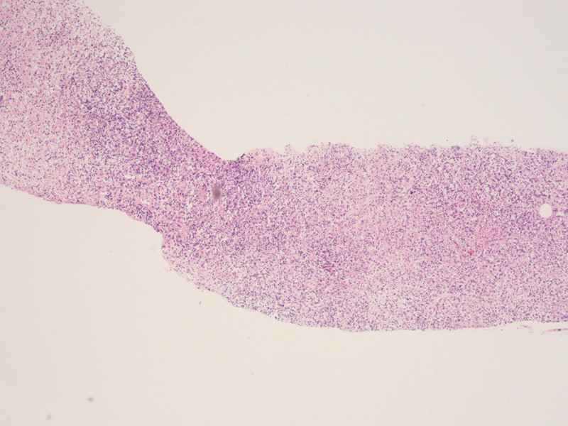
Diagnosis: A US-guided biopsy was conducted with the final pathological diagnosis of PTLD (polymorphic).
Interventions: After receiving 3 times of rituximab and symptomatic treatment, blood creatinine returned to normal but the mass was still progressing in the patient. Therefore, the treatment approach was modified to immune-chemotherapy.
Outcomes: The patient was in a stable condition to date.
Lessons: PTLD is a rare complication in a transplanted kidney. US and CEUS are the preferred imaging methods in renal transplant patients due to their good repeatability and no nephrotoxicity. This case demonstrates that continuous dynamic monitoring by using US and CEUS has significant value in the detection and diagnosis of PTLD in a transplanted kidney, suggesting early clinical intervention to avoid further progression.
Copyright © 2024 the Author(s). Published by Wolters Kluwer Health, Inc.
Conflict of interest statement
The authors have no conflicts of interest to disclose.
Figures

Similar articles
-
Application of contrast-enhanced ultrasonography in the diagnosis of post-kidney transplant lymphoproliferative disorder in native kidney- a case report.BMC Cancer. 2019 Nov 21;19(1):1135. doi: 10.1186/s12885-019-6355-0. BMC Cancer. 2019. PMID: 31752767 Free PMC article.
-
Multimodality imaging features, treatment, and prognosis of post-transplant lymphoproliferative disorder in renal allografts: A case report and literature review.Medicine (Baltimore). 2018 Apr;97(17):e0531. doi: 10.1097/MD.0000000000010531. Medicine (Baltimore). 2018. PMID: 29703027 Free PMC article.
-
Application of contrast-enhanced ultrasound in the diagnosis of post-transplant lymphoproliferative disease after hematopoietic stem cell transplantation: A case report.Medicine (Baltimore). 2021 Jan 15;100(2):e24047. doi: 10.1097/MD.0000000000024047. Medicine (Baltimore). 2021. PMID: 33466157 Free PMC article.
-
Contrast-enhanced ultrasound findings of post-transplant lymphoproliferative disorder in a transplanted kidney: A case report and literature review.J Radiol Case Rep. 2015 Oct 31;9(10):26-34. doi: 10.3941/jrcr.v9i10.2602. eCollection 2015 Oct. J Radiol Case Rep. 2015. PMID: 26629291 Free PMC article. Review.
-
Lymphoproliferative disorders after renal transplantation: role of medical imaging.Eur Radiol. 1998;8(9):1686-93. doi: 10.1007/s003300050614. Eur Radiol. 1998. PMID: 9866789 Review.
Cited by
-
Imaging misdiagnosis of urothelial carcinoma of the kidney graft as a post-transplant lymphoproliferative disorder: a case description.Quant Imaging Med Surg. 2025 Jan 2;15(1):1073-1079. doi: 10.21037/qims-24-1323. Epub 2024 Dec 27. Quant Imaging Med Surg. 2025. PMID: 39838984 Free PMC article. No abstract available.
References
-
- Yaginuma T, Yamamoto H, Mitome J, et al. . Successful treatment of monomorphic primary central nervous system post-transplantation lymphoproliferative disorder 5 years after kidney transplantation. Transpl Infect Dis. 2012;14:E102–106. - PubMed
-
- Opelz G, Dohler B. Lymphomas after solid organ transplantation: a collaborative transplant study report. Am J Transplant. 2004;4:222–30. - PubMed
Publication types
MeSH terms
Substances
LinkOut - more resources
Full Text Sources
Medical
Research Materials
