. 2024 Jun 4;23(6):836-853.
doi: 10.1158/1535-7163.MCT-23-0868.

Structural Basis for Multivalent MUC16 Recognition and Robust Anti-Pancreatic Cancer Activity of Humanized Antibody AR9.6

Affiliations

Structural Basis for Multivalent MUC16 Recognition and Robust Anti-Pancreatic Cancer Activity of Humanized Antibody AR9.6

Eric N Aguilar et al. Mol Cancer Ther. .

Abstract

Mucin-16 (MUC16) is a target for antibody-mediated immunotherapy in pancreatic ductal adenocarcinoma (PDAC) among other malignancies. The MUC16-specific monoclonal antibody AR9.6 has shown promise for PDAC immunotherapy and imaging. Here, we report the structural and biological characterization of the humanized AR9.6 antibody (huAR9.6). The structure of huAR9.6 was determined in complex with a MUC16 SEA (Sea urchin sperm, Enterokinase, Agrin) domain. Binding of huAR9.6 to recombinant, shed, and cell-surface MUC16 was characterized, and anti-PDAC activity was evaluated in vitro and in vivo. HuAR9.6 bound a discontinuous, SEA domain epitope with an overall affinity of 88 nmol/L. Binding affinity depended on the specific SEA domain(s) present, and glycosylation modestly enhanced affinity driven by favorable entropy and enthalpy and via distinct transition state thermodynamic pathways. Treatment with huAR9.6 reduced the in vitro growth, migration, invasion, and clonogenicity of MUC16-positive PDAC cells and patient-derived organoids (PDO). HuAR9.6 blocked MUC16-mediated ErbB and AKT activation in PDAC cells, PDOs, and patient-derived xenografts and induced antibody-dependent cellular cytotoxicity and complement-dependent cytotoxicity. More importantly, huAR9.6 treatment caused substantial PDAC regression in subcutaneous and orthotopic tumor models. The mechanism of action of huAR9.6 may depend on dense avid binding to homologous SEA domains on MUC16. The results of this study validate the translational therapeutic potential of huAR9.6 against MUC16-positive PDACs.


Conflict of interest statement

Conflict of interest statement

C.L. Brooks received grants from Quest PharmaTech Inc. (AR9.6 patent assignee, US-11773183-B2) during the conduct of the study and grants from Quest PharmaTech Inc. P. Radhakrishnan and M.A. Hollingsworth are inventors on a patent related to huAR9.6 (US-11773183-B2). All other authors declare no competing interests.

Figures

Figure 1.
Figure 1.. Structural and biophysical characterization of huAR9.6.
(A) X-ray structure of murine AR9.6 (mAR9.6, cyan/green) and humanized AR9.6 (huAR9.6, magenta/salmon). (B) Structural alignment of mAR9.6 (cyan/green) and huAR9.6 (magenta/salmon). The variable domains (VH, VL) have the highest degree of similarity, the constant domains (CH, CL) are also similar. (C) MUC16 SEA5 constructs for huAR9.6 epitope mapping. 1.2 copies of a tandem repeat (1.2TR), the SEA5 domain with N-terminal or C-terminal flanking sequences rich in PST, PST-SEA5, SEA5-PST, the SEA domain alone, and overlapping 20 residue peptides covering the SEA5 domain were produced in E. coli as C-terminal Trx fusions. (D) Epitope mapping of huAR9.6 by indirect ELISA. SEA5 domain containing proteins were immobilized and detected with two concentrations of huAR9.6. The minimum epitope was an intact SEA5 domain. (E) Indirect ELISA and apparent affinity (Kapp) of huAR9.6 IgG. The mAb bound with equal Kapp to all constructs containing an intact SEA5 domain (1.2TR Kapp = 0.37 nM, PST-SEA5 Kapp = 0.25 nM, SEA5 Kapp = 0.40 nM, SEA5-PST Kapp = 0.42 nM). (F) Temperature dependence of KD and van’t Hoff approximation to calculate thermodynamic parameters of huAR9.6 binding to glycosylated and aglycosylated MUC16 domains. (G) Temperature dependance of kon and koff used to calculate transition state thermodynamic parameters using the Eyring approximation. (H) Energy diagram of the association and dissociation of huAR9.6 with glycosylated and aglycosylated MUC16 domains. A+B represents the energy of the Fab-antigen unbound state, A-B is the transition state of association, AB is the Fab-antigen bound state, A-B* represents the transition state of dissociation, and A+B* is the dissociated state. Error bars in all graphs represent the SD of n=3 replicates.
Figure 2.
Figure 2.. Epitope mapping of huAR9.6 by HDX-MS and X-ray crystallography.
(A) HDX-MS heat map showing the difference in SEA5 deuterium uptake following interaction with mAR9.6 Fab and huAR9.6 IgG. Five regions were identified that displayed a significant increase in deuterium protection (B) Regions of possible SEA5 interaction with AR9.6 identified by HDX-MS. (C) Co-crystal structure of mAR9.6 Fab fragment (ribbon diagram, light chain shown in cyan, heavy chain shown in green) in complex with SEA5 (yellow surface). The interaction surface with the antibody light chain (white) is significantly smaller than with the heavy chain (maroon). (D) Summary of polar interactions between AR9.6 light chain (cyan) and heavy chain (green) with SEA5 (yellow). (E) AR9.6 epitope (red) on SEA5 (yellow) as determined by X-ray crystallography. (F) Simple model of glycosylated SEA5 with N-linked glycans (yellow) an O-linked glycans (purple) docked with AR9.6 (heavy chain shown in green, light chain shown in cyan). The antibody may interact with both O-linked and N-linked glycans on the antigen.
Figure 3.
Figure 3.. HuAR9.6 demonstrates robust anti-PDAC activity.
Cell viability assays using alamarBlue in (A) T3M4 and (B) Capan-1 cells treated with vehicle (PBS), isotype control huIgG and huAR9.6 at increasing concentrations (n=5). Quantification as percent wound closure showing cell migration upon vehicle (PBS), isotype control huIgG (40 μg/mL) and huAR9.6 (40 μg/mL) treatment in (C) T3M4 and (D) Capan-1 cells (n=3). Transwell invasion and its quantification assays showing vehicle (PBS), isotype control huIgG and huAR9.6 (40 μg/mL) treated (E and F) T3M4 and (G and H) Capan-1 cells (n=3). Colony formation and its quantification assay assessing clonogenicity of (I and J) T3M4 and (K and L) Capan-1 cells treated with vehicle (PBS), isotype control huIgG (40 μg/mL) and huAR9.6 (40 μg/mL). Mean ± SD (n=3 replicates), p < 0.05 was considered as statistically significant using one-way ANOVA with Tukey’s multiple comparisons.
Figure 4.
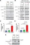
Figure 4.. huAR9.6 disrupts the ErbB signaling axis and induces apoptotic cell death.
(A) Western blotting for p-ErbB3 (Y1289), ErbB3, pErbB1 (Y1173), ErbB1, pFAK (Y397), FAK, pAKT (S473), AKT in T3M4 and Capan-1 cells treated with vehicle (PBS), isotype control huIgG (40 μg/mL) and huAR9.6 (40 μg/mL). (B) Western blotting for p-ErbB3 (Y1289), ErbB3, pErbB1 (Y1173), ErbB1, pFAK (Y397), FAK, pAKT (S473), AKT in PDX derived cells treated with vehicle (PBS), isotype control huIgG (40 μg/mL) and huAR9.6 (40 μg/mL). Quantification of % apoptotic cells in (C) T3M4 and (D) Capan-1 cells treated with vehicle (PBS), isotype control huIgG and huAR9.6 (n=3). (E) Western blotting for PARP, cleaved caspase 3 (D175), caspase 3 in T3M4 and Capan-1 cells treated with vehicle (PBS), isotype control huIgG (40 μg/mL) and huAR9.6 (40 μg/mL). Loading control: β-Actin. Mean ± SD, p < 0.05 was considered as statistically significant using one-way ANOVA with Tukey’s multiple comparisons.
Figure 5.
Figure 5.. huAR9.6 reduces tumor burden in models of PDAC.
(A) T3M4 sub-cutaneous tumor-bearing animals treated with vehicle and huAR9.6 (500 μg/25 g bodyweight, i.p. every 72 h). Tumor volume measurements in mm3 on alternate days starting from day of treatment administration (day 9). (B) Tumor weight measurements in grams at the experimental endpoint (day 19). Quantification of (C) Ki67 and (D) CD31 positive nuclei depicted as percent of vehicle treated tumors (n=5). T3M4 orthotopic tumor-bearing animals treated with vehicle (PBS), isotype control huIgG and huAR9.6 (500 μg/25 g bodyweight, i.p.) for 4 doses. At the experimental endpoint (day 28), mice were euthanized. (E) Tumor volume (mm3) and (F) Tumor weight (grams) at the experimental endpoint (day 28). (G) H&E and Ki67 staining on PDOs treated with vehicle (PBS), isotype control huIgG and huAR9.6 (40 μg/mL for 24 h). Scale bar 50 μm. (H) Quantification of Ki67 positive nuclei depicted as percent of vehicle treated tumors (n=5 fields). (I) Diagrammatic representation of ADCC and CDC potentiated by huAR9.6 after binding to MUC16 on tumor cells (target cells). (J) Quantification of ADCC as % dead target (tumor) cells (n=3). (K) Quantification of CDC as % of dead cells (n=3). Mean ± SD, p < 0.05 was considered as statistically significant using the Student’s t-test or one-way ANOVA with Tukey’s multiple comparisons.

References

    1. Siegel RL, Miller KD, Wagle NS, Jemal A. Cancer statistics, 2023. CA Cancer J Clin. United States; 2023;73:17–48. - PubMed
    1. Aithal A, Rauth S, Kshirsagar P, Shah A, Lakshmanan I, Junker WM, et al. MUC16 as a novel target for cancer therapy. Expert Opin Ther Targets. 2018;22:675–86. - PMC - PubMed
    1. Marcos-Silva L, Narimatsu Y, Halim A, Campos D, Yang Z, Tarp MA, et al. Characterization of binding epitopes of CA125 monoclonal antibodies. J Proteome Res. United States; 2014;13:3349–59. - PubMed
    1. Thomas D, Sagar S, Liu X, Lee H-R, Grunkemeyer JA, Grandgenett PM, et al. Isoforms of MUC16 activate oncogenic signaling through EGF receptors to enhance the progression of pancreatic cancer. Mol Ther. 2021;29:1557–71. - PMC - PubMed
    1. Muniyan S, Haridas D, Chugh S, Rachagani S, Lakshmanan I, Gupta S, et al. MUC16 contributes to the metastasis of pancreatic ductal adenocarcinoma through focal adhesion mediated signaling mechanism. Genes Cancer. 2016;7:110–24. - PMC - PubMed

MeSH terms