Review
. 2024 May 7;120(6):567-580.
doi: 10.1093/cvr/cvae044.

Cardiac and perivascular myofibroblasts, matrifibrocytes, and immune fibrocytes in hypertension; commonalities and differences with other cardiovascular diseases

Affiliations
Review

Cardiac and perivascular myofibroblasts, matrifibrocytes, and immune fibrocytes in hypertension; commonalities and differences with other cardiovascular diseases

Keiichi Torimoto et al. Cardiovasc Res. .

Abstract

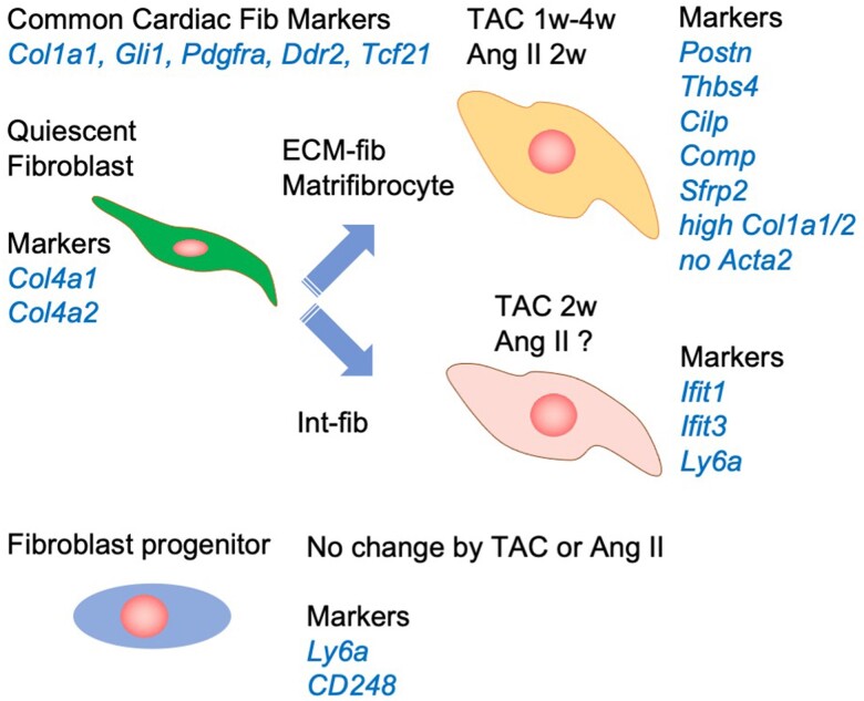
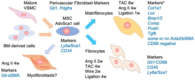
Hypertension is a major cause of cardiovascular diseases such as myocardial infarction and stroke. Cardiovascular fibrosis occurs with hypertension and contributes to vascular resistance, aortic stiffness, and cardiac hypertrophy. However, the molecular mechanisms leading to fibroblast activation in hypertension remain largely unknown. There are two types of fibrosis: replacement fibrosis and reactive fibrosis. Replacement fibrosis occurs in response to the loss of viable tissue to form a scar. Reactive fibrosis occurs in response to an increase in mechanical and neurohormonal stress. Although both types of fibrosis are considered adaptive processes, they become maladaptive when the tissue loss is too large, or the stress persists. Myofibroblasts represent a subpopulation of activated fibroblasts that have gained contractile function to promote wound healing. Therefore, myofibroblasts are a critical cell type that promotes replacement fibrosis. Although myofibroblasts were recognized as the fibroblasts participating in reactive fibrosis, recent experimental evidence indicated there are distinct fibroblast populations in cardiovascular reactive fibrosis. Accordingly, we will discuss the updated definition of fibroblast subpopulations, the regulatory mechanisms, and their potential roles in cardiovascular pathophysiology utilizing new knowledge from various lineage tracing and single-cell RNA sequencing studies. Among the fibroblast subpopulations, we will highlight the novel roles of matrifibrocytes and immune fibrocytes in cardiovascular fibrosis including experimental models of hypertension, pressure overload, myocardial infarction, atherosclerosis, aortic aneurysm, and nephrosclerosis. Exploration into the molecular mechanisms involved in the differentiation and activation of those fibroblast subpopulations may lead to novel treatments for end-organ damage associated with hypertension and other cardiovascular diseases.

Keywords: Fibroblast; Fibrosis; Hypertension; Myocardial Infarction; Pressure overload.


Conflict of interest statement

Conflict of interest: None declared.

Figures

Figure 1
Figure 1
Fibroblast subpopulations and their markers seen upon MI. (A) Simplified 4 fibroblast stage proposal in response to MI. Around days 2∼4 post MI, proliferation and migration of fibroblast populations are observed. ECM production is not enhanced. These proliferating fibroblasts expressing periostin will differentiate to myofibroblasts at the infract zone on day 4∼7. Myofibroblasts are the main fibroblast population contributing to the cardiac scar formation. These fibroblasts gain contractility and actively produce ECM components to complete replacement fibrosis. Upon completion of the scar, myofibroblasts are believed to further differentiate to matrifibrocytes to maintain the scar. However, cardiac mechanical stress and neurohormonal stress persist due to cardiac dysfunction caused by MI and insufficient compensation by the scar, leading to reactive fibrosis. Matrifibrocytes produce ECM components and contribute to reactive fibrosis as an adaptation to chronic cardiac injury. Marker genes and proteins preferentially expressed in each stage have been reported. Resting fibroblasts can be identified by the fibroblast markers as discussed. Proliferating fibroblasts expressing stem cell markers including Ly6A/Sca1, activated fibroblast marker periostin, cell cycle genes, inflammatory cytokines, and chemokines demonstrate their pro-fibrotic pro-inflammatory roles in the initiation of cardiac inflammation and fibrosis. Both myofibroblasts and matrifibrocytes highly express ECM genes and are involved in early replacement fibrosis and chronic reactive fibrosis, respectively. Myofibroblasts express periostin and contractile genes including Acta2/αSMA. Matrifibrocytes localize to scar site and border zones and express genes related to bone and cartilage remodelling, such as Comp, Chad, and Cilp1/2. Italics indicate the marker genes. (B) Updated understanding of fibroblast subpopulations and their markers seen upon MI. Resident resting fibroblasts appear to be the major source of fibroblasts involved in scar formation and reactive fibrosis upon MI. Three IR fibroblasts are the first responders to MI and cause an acute pro-inflammatory response at the site. Myofibroblast subpopulations proliferate and are actively involved in matrix production, scar formation and inflammatory conditions. At days 14∼28, these fibroblasts differentiate to matrifibrocytes, late resolution fibroblasts, and fibrocytes. These cell types play major roles in chronic interstitial fibrosis and inflammation as adaptation to post MI cardiac dysfunction. It is also important to note that some matrifibrocytes and other late stage fibroblasts may show stress-induced senescence and senescence-associated secretory phenotype/SASP, thus accelerate cardiac remodelling. Abbreviations are Act-fib; activated fibroblast; fib; fibroblast, Myofib; myofibroblast. Italics indicate the marker genes.
Figure 2
Figure 2
Cardiac fibroblast subpopulations and their markers seen upon TAC or Ang II infusion. Significant increases in Col1a1/2 and Postn ECM-Fib occur days 14 and 28 post TAC. This population appears to represent Fibroblast-Cilp plus Fibroblast-Thbs4 populations seen upon Ang II infusion as well as matrifibrocytes upon MI. ECM-Fib cells do not express myofibroblast marker, Acta2. These fibroblasts are therefore the main mediator of cardiac interstitial fibrosis in response to enhanced afterload (TAC or Ang II hypertension). In addition, the transient increase in Interferon-Fib cells at 2 weeks post TAC suggests the role to be pro-inflammatory. The inflammatory fibroblasts express stem cell marker, Ly6A but do not proliferate. Fibroblast progenitor cells are also present with no change in the population upon TAC or Ang II infusion. Abbreviations are Ang II, angiotensin II; col, collagen; ECM-Fib, extracellular matrix fibroblasts; Int-fib, interferon-; TAC, Transverse aortic constriction. Italics indicate the marker genes.
Figure 3
Figure 3
Current understanding of fibroblast cell types involved in perivascular fibrosis induced by mechanical and neurohormonal stress. Matrifibrocytes and fibrocytes are two major subtypes identified as contributing to perivascular fibrosis and vascular inflammation. The major collagen producing matrifibrocytes seen in vascular adventitia may be similar to those found in the cardiac fibroblast populations. Resident MSC is the source of these activated fibroblasts. The perivascular MSC may have mature VSMC and BM cell lineage. Fibrocytes may have similar roles as interferon fibroblasts seen in the heart upon TAC or Ang II infusion. Again, the contribution of myofibroblasts to perivascular fibrosis seems relatively limited. Abbreviations: αSMA, smooth muscle α actin; AdvSca1, adventitial Sca1; Ang II, angiotensin II; BM, bone marrow; Fib, fibroblast; MSC, mesenchymal stem cell; Sca1, stem cell antigen 1; TAC, Transverse aortic constriction; VSMC, vascular smooth muscle cell. Italics indicate the marker genes.

References

    1. Mills KT, Stefanescu A, He J. The global epidemiology of hypertension. Nat Rev Nephrol 2020;16:223–237. - PMC - PubMed
    1. Al-Makki A, DiPette D, Whelton PK, Murad MH, Mustafa RA, Acharya S, Beheiry HM, Champagne B, Connell K, Cooney MT, Ezeigwe N, Gaziano TA, Gidio A, Lopez-Jaramillo P, Khan UI, Kumarapeli V, Moran AE, Silwimba MM, Rayner B, Sukonthasan A, Yu J, Saraffzadegan N, Reddy KS, Khan T. Hypertension pharmacological treatment in adults: a world health organization guideline executive summary. Hypertension 2022;79:293–301. - PMC - PubMed
    1. Frangogiannis NG. Cardiac fibrosis. Cardiovasc Res 2021;117:1450–1488. - PMC - PubMed
    1. Humeres C, Frangogiannis NG. Fibroblasts in the infarcted, remodeling, and failing heart. JACC Basic Transl Sci 2019;4:449–467. - PMC - PubMed
    1. Oka T, Xu J, Kaiser RA, Melendez J, Hambleton M, Sargent MA, Lorts A, Brunskill EW, Dorn GW II, Conway SJ, Aronow BJ, Robbins J, Molkentin JD. Genetic manipulation of periostin expression reveals a role in cardiac hypertrophy and ventricular remodeling. Circ Res 2007;101:313–321. - PMC - PubMed

Publication types