Review
. 2024 Sep;34(9):707-727.
doi: 10.1016/j.tcb.2024.01.009. Epub 2024 Feb 22.

Chromosome compartmentalization: causes, changes, consequences, and conundrums

Affiliations
Review

Chromosome compartmentalization: causes, changes, consequences, and conundrums

Heng Li et al. Trends Cell Biol. 2024 Sep.

Abstract

The spatial segregation of the genome into compartments is a major feature of 3D genome organization. New data on mammalian chromosome organization across different conditions reveal important information about how and why these compartments form and change. A combination of epigenetic state, nuclear body tethering, physical forces, gene expression, and replication timing (RT) can all influence the establishment and alteration of chromosome compartments. We review the causes and implications of genomic regions undergoing a 'compartment switch' that changes their physical associations and spatial location in the nucleus. About 20-30% of genomic regions change compartment during cell differentiation or cancer progression, whereas alterations in response to a stimulus within a cell type are usually much more limited. However, even a change in 1-2% of genomic bins may have biologically relevant implications. Finally, we review the effects of compartment changes on gene regulation, DNA damage repair, replication, and the physical state of the cell.

Keywords: 3D genome; cell differentiation; chromosome compartments; epigenetics; gene regulation.


Conflict of interest statement

Declaration of interests R.P.M. is a member of the Cell Press Statistical Advisory Board. The other authors declare no conflicts of interest.

Figures
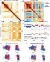
Figure 1.
Figure 1.. Detecting compartments using Hi-C and chromatin tracing.
A) Compartmentalization is visible as plaid patterns of interaction in contact maps from IMR90 (data from [5]) both within chr21 (top) and between chr21 and chr22 (bottom). B) Calculating the distance-normalized correlation map emphasizes the compartment pattern. C) A comparison of two-state (A/B, 1st row), Rao subcompartment [5] (2nd row), and deGeco [13] 4-state (3rd row) classification of compartments. Genomic positions along each track correspond to the IMR90 chr21 positions in the correlation map in panel (B). A1/A2 and S1/S2 generally overlap with the A compartment while B1/B2 and S3/S4 overlap with the B compartment. Bottom row: SPRITE can capture multi-way contacts that can represent different ways compartments interact in different cells. D) and E) Chromatin tracing data from two different IMR90 cells (from [128]) shows the position of chromosome regions (beads) in the 3D space of the nucleus. Beads are colored by PCA-derived A/B compartment assignment (D) or Rao subcompartment (E) to show that these regions are indeed spatially segregated in individual cells. Bottom row shows a rotated view of the same structure.
Figure 2.
Figure 2.. Epigenomic features typically associated with the A and B compartment.
A cartoon showing simplified typical correlation patterns between A/B compartments (Hi-C PC1), transcriptional levels, chromatin accessibility, and ChIP-seq signals for nuclear lamina association (lamin A/C and lamin B) and five key histone modifications (active marks H3K27ac, H3K4me1, H3K36me3 and repressive marks H3K9me3 and H3K27me3), and replication timing (green=early, cyan=late).
Figure 3.
Figure 3.. What conditions can change compartmentalization?
The most dramatic compartment changes are typically observed during differentiation and reprogramming or cancer progression (orange). Exogenous stimuli such as infection/inflammation, DNA damaging agents, or hexanediol treatment and internal protein changes such as lamin mutations can lead to minor to moderate compartment changes (blue). Other extrinsic factors (hypotonic swelling, heatshock) or protein alterations (CTCF removal) have very little to no effect on compartment identity (grey) (see Table 1 for specific citations and examples). Figure created using BioRender.
Figure 4.
Figure 4.. Factors influencing whether an epigenetic change will cause compartment switch.
Genomic regions in the A compartment possess characteristics that make them less likely (Region 1) or more likely (Region 2) to switch to the B compartment after targeted epigenetic changes. Region 1 is spatially far from the B compartment, gene dense with high transcription, and has many active chromatin marks. Region 2, in contrast, has only moderate gene expression, intermediate histone modifications, and is proximal to the B compartment. The switch to B is also more likely if there is a high level of expression of heterochromatin-associated proteins (represented by yellow ovals).
Figure 5.
Figure 5.. Types of changes in A/B compartmentalization.
Compartments can change either by an internal region of a compartment flipping out to associate with the opposite compartment (A) or by boundary shifting on one or both sides (B). Left panels: a cartoon of what this type of compartment change could look like in the path of the chromosome (red = A, blue = B). Middle panels: what the PCA quantification of this type of compartment change looks like. Right panels: Examples of these types of compartment changes from real data. Top: flipping out observed in a comparison of different types of breast cancer (adapted with permission from: Kim et al., 2022 [37]). Bottom: boundary shifting observed in the differentiation from ESC to cardiomyocyte (adapted with permission from: Bertero et al., 2019 [27])
Figure 6.
Figure 6.. Coordinated changes in A/B compartments, gene expression, and replication timing in differentiating cells.
Cell differentiation is accompanied by rearrangement of 3D chromatin structure, chromatin modification and gene regulation. The shift of a chromosome region from B to A compartment (top panel to bottom panel) coincides with changes in replication timing from late (green) to early (yellow), shifts in histone modifications (see dotted zoom inset) and increases in expression of genes in the region.

References

    1. Caspersson T et al. (1970) Differential binding of alkylating fluorochromes in human chromosomes. Experimental Cell Research 60 (3), 315–319. - PubMed
    1. Littau VC et al. (1964) ACTIVE AND INACTIVE REGIONS OF NUCLEAR CHROMATIN AS REVEALED BY ELECTRON MICROSCOPE AUTORADIOGRAPHY*. Proceedings of the National Academy of Sciences 52 (1), 93–100. - PMC - PubMed
    1. Lieberman-Aiden E et al. (2009) Comprehensive mapping of long-range interactions reveals folding principles of the human genome. science 326 (5950), 289–293. - PMC - PubMed
    1. Mirny LA et al. (2019) Two major mechanisms of chromosome organization. Curr Opin Cell Biol 58, 142–152. - PMC - PubMed
    1. Rao SS et al. (2014) A 3D map of the human genome at kilobase resolution reveals principles of chromatin looping. Cell 159 (7), 1665–80. - PMC - PubMed

LinkOut - more resources