. 2024 Feb 23;12(1):32.
doi: 10.1186/s40478-024-01742-x.

Stimulating VAPB-PTPIP51 ER-mitochondria tethering corrects FTD/ALS mutant TDP43 linked Ca2+ and synaptic defects

Affiliations

Stimulating VAPB-PTPIP51 ER-mitochondria tethering corrects FTD/ALS mutant TDP43 linked Ca2+ and synaptic defects

Andrea Markovinovic et al. Acta Neuropathol Commun. .

Abstract

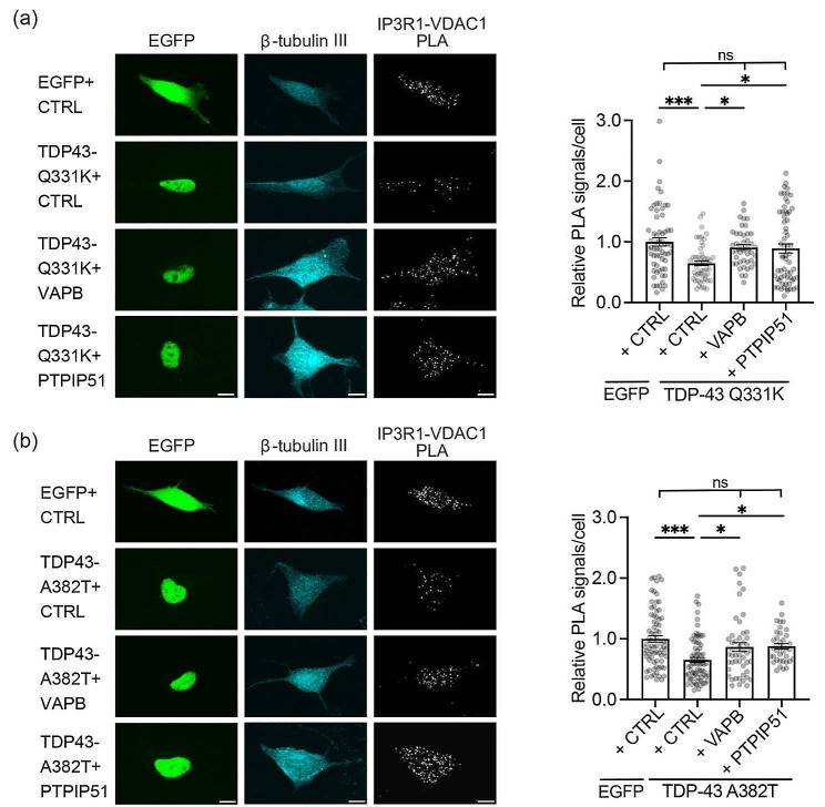
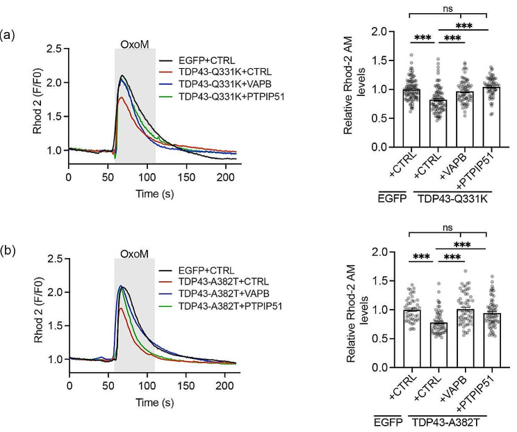
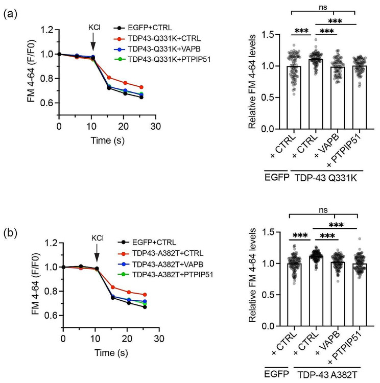
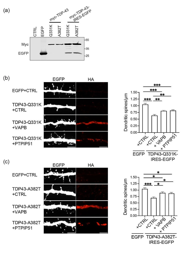
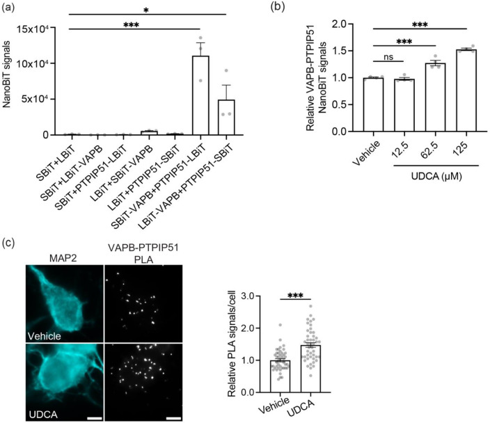
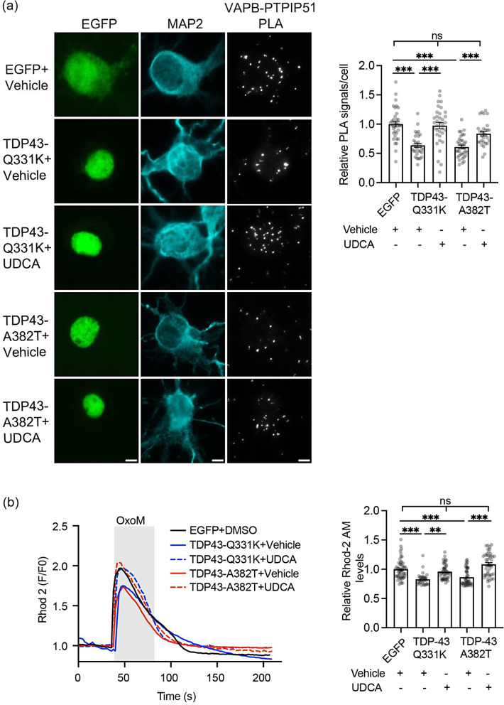
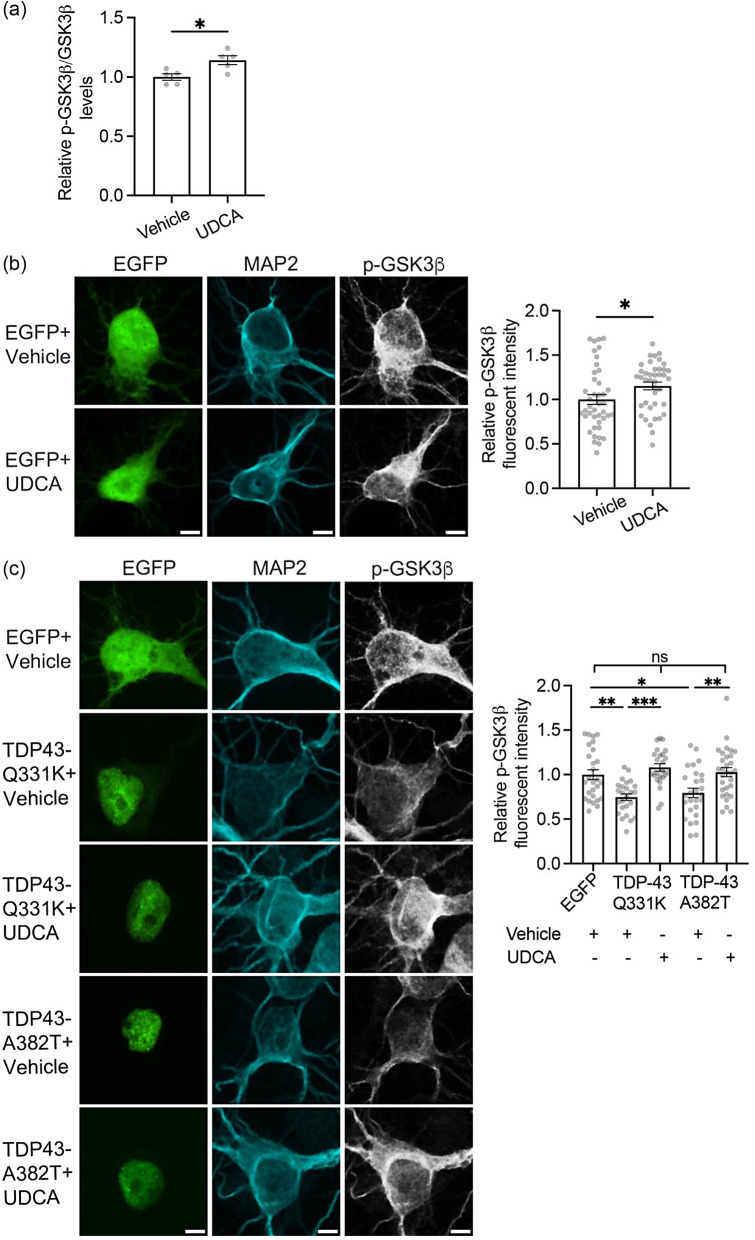
Frontotemporal dementia (FTD) and amyotrophic lateral sclerosis (ALS) are clinically linked major neurodegenerative diseases. Notably, TAR DNA-binding protein-43 (TDP43) accumulations are hallmark pathologies of FTD/ALS and mutations in the gene encoding TDP43 cause familial FTD/ALS. There are no cures for FTD/ALS. FTD/ALS display damage to a broad range of physiological functions, many of which are regulated by signaling between the endoplasmic reticulum (ER) and mitochondria. This signaling is mediated by the VAPB-PTPIP51 tethering proteins that serve to recruit regions of ER to the mitochondrial surface so as to facilitate inter-organelle communications. Several studies have now shown that disrupted ER-mitochondria signaling including breaking of the VAPB-PTPIP51 tethers are features of FTD/ALS and that for TDP43 and other familial genetic FTD/ALS insults, this involves activation of glycogen kinase-3β (GSK3β). Such findings have prompted suggestions that correcting damage to ER-mitochondria signaling and the VAPB-PTPIP51 interaction may be broadly therapeutic. Here we provide evidence to support this notion. We show that overexpression of VAPB or PTPIP51 to enhance ER-mitochondria signaling corrects mutant TDP43 induced damage to inositol 1,4,5-trisphosphate (IP3) receptor delivery of Ca2+ to mitochondria which is a primary function of the VAPB-PTPIP51 tethers, and to synaptic function. Moreover, we show that ursodeoxycholic acid (UDCA), an FDA approved drug linked to FTD/ALS and other neurodegenerative diseases therapy and whose precise therapeutic target is unclear, corrects TDP43 linked damage to the VAPB-PTPIP51 interaction. We also show that this effect involves inhibition of TDP43 mediated activation of GSK3β. Thus, correcting damage to the VAPB-PTPIP51 tethers may have therapeutic value for FTD/ALS and other age-related neurodegenerative diseases.

Keywords: Alzheimer’s disease; Amyotrophic lateral sclerosis; Frontotemporal dementia; Neurodegenerative diseases; Parkinson’s disease; TDP43.


Conflict of interest statement

The authors declare they have no competing interests.

Figures

Fig. 1
Fig. 1
Expression of VAPB or PTPIP51 rescues mutant TDP43 induced disruption to the IP3 receptor-VDAC1 interaction. (a and b) Representative images of IP3 receptor type1-VDAC1 PLAs in SH-SY5Y cells transfected with EGFP + control (CTRL) vector, EGFP-tagged-TDP43-Q331K or -TDP43-A382T + control vector and EGFP-tagged TDP43-Q331K/EGFP-TDP43-A382T + either VAPB or PTPIP51 as indicated. TDP43 was identified via the EGFP tag and cells immunostained for β-tubulin III (artificially shown in cyan). Scale bars = 10 μm. Bar charts show PLA signals per cell after normalisation to control EGFP + CTRL cell data. Data were analysed by one-way ANOVA with Tukey’s post hoc test; (a) N = 43–63 cells from 3 independent experiments, (b) N = 38–75 cells from 3 independent experiments. Error bars are SEM; * p≤0.05, *** p≤0.001, ns not significant
Fig. 2
Fig. 2
Expression of VAPB or PTPIP51 rescues mutant TDP43 induced disruption to IP3 receptor mediated delivery of Ca2+ to mitochondria. (a and b) SH-SY5Y cells were transfected with EGFP + control (CTRL) vector, EGFP-TDP43-Q331K or TDP43-A382T + control vector and EGFP-TDP43-Q331K/EGFP-TDP43-A382T + either VAPB or PTPIP51 as indicated. Cells were loaded with Rhod-2 AM and Ca2+ release induced by treatment with oxotremorine-M (OxoM). Representative Rhod-2 AM fluorescence traces are shown on the left with OxoM treatment depicted by shaded area; normalised peak values are shown in the bar charts on the right. Expression of TDP43-Q331K or TDP43-A382T reduced mitochondrial Ca2+ levels and this was rescued by VAPB/PTPIP51 expression. Data were analysed by one-way ANOVA with Tukey’s post hoc test. N = 62–96 cells from 3 independent experiments in (a) and N = 48–69 cells from 3 independent experiments in (b). Error bars are SEM; *** p≤0.001, ns not significant
Fig. 3
Fig. 3
Expression of VAPB or PTPIP51 rescues mutant TDP43 induced disruption to synaptic vesicle release. (a and b) Kinetics of FM 4–64 release from synaptic boutons in cortical neurons transfected with (a) EGFP + CTRL, EGFP + TDP43-Q331K + CTRL or EGFP + TDP43-Q331K + either VAPB or PTPIP51 and (b) EGFP + CTRL, EGFP + TDP43-A382T + CTRL or EGFP + TDP43-A382T + either VAPB or PTPIP51 as indicated. Synaptic vesicle release was induced by treatment with KCl and FM 4–64 signals were determined from images acquired by time-lapse microscopy. Representative FM 4–64 traces are shown on the left; bar charts show F/F0 FM 4–64 fluorescent signals 5 s post KCl treatment. Data were analysed by one-way ANOVA with Tukey’s post hoc test. N = 68–102 boutons in (a) and N = 133–146 boutons in (b) from 3 independent experiments. Error bars are SEM; *** p≤0.001, ns not significant
Fig. 4
Fig. 4
Expression of VAPB or PTPIP51 rescues mutant TDP43 induced disruption to dendritic spine numbers. (a) Characterisation of mutant TDP43-IRES-EGFP lentivirus. HEK293 cells were transfected with control vector (CTRL), EGFP, Myc-TDP43-Q331K or Myc-TDP43-A382T as negative and positive controls, and samples run on SDS-PAGE alongside samples of neurons infected with lentivirus expressing Myc-tagged TDP43-Q331K-IRES-EGFP or TDP43-A382T-IRES-EGFP as indicated. Samples were probed on immunoblots for TDP43 via the Myc tags and EGFP; molecular masses are shown on the right. (b and c) Representative super resolution SIM images of dendritic spines in neurons treated with lentivirus expressing (b) EGFP + CTRL, TDP43-Q331K-IRES-EGFP + CTRL, TDP43-Q331K-IRES-EGFP + HA tagged VAPB or TDP43-Q331K-IRES-EGFP + HA tagged PTPIP51 or (c) EGFP + CTRL, TDP43-A383T-IRES-EGFP + CTRL, TDP43-A382T-IRES-EGFP + HA tagged VAPB or TDP43-A382T-IRES EGFP + HA tagged PTPIP51. Neurons were infected with lentivirus at DIV 12 and analysed at DIV 15. Exogenous VAPB and PTPIP51 were identified by immunostaining for the HA tags. Scale bars = 5 μm. Bar charts show spine densities calculated as described (spines/µm) [17]. Data were analysed by one-way ANOVA with Tukey’s post hoc test. N = 20–26 neurons from 3 independent experiments; error bars are SEM. * p≤0.05, ** p≤0.01, *** p≤0.001
Fig. 5
Fig. 5
UDCA stimulates the VAPB-PTPIP51 interaction. (a) Characterisation of VAPB-PTPIP51 NanoBiT assays. SH-SY5Y cells were transfected with either empty Small and Large BiT plasmids (SBiT + LBiT), SBiT + LBiT-VAPB, SBiT + LBiT-PTPIP51, LBiT + SBiT-VAPB, LBiT + PTPIP51-SBiT, SBiT-VAPB + PTPIP51-LBiT or LBiT-VAPB + PTPIP51-SBiT as indicated. Signals above background were only detected in cells transfected with SBiT-VAPB + PTPIP51-LBiT or LBiT-VAPB + PTPIP51-SBiT and the highest signals were detected in SBiT-VAPB + PTPIP51-LBiT cells. Data were analysed by one-way ANOVA with Tukey’s post hoc test. Data are from 3 independent experiments each with N = 6; error bars are SEM. * p≤0.05, *** p≤0.001 (b) UDCA stimulates VAPB-PTPIP51 binding in NanoBiT assays. SH-SY5Y cells were transfected with SBiT-VAPB + PTPIP51-LBiT and then treated with vehicle or increasing concentrations of UDCA for 24 h. 62.5 µM and 125 µM UDCA both stimulated VAPB-PTPIP51 binding. Data were analysed by one-way ANOVA with Tukey’s post hoc test. Data are from 4 independent experiments each with N = 6. Error bars are SEM. *** p≤0.001, ns not significant. (c) UDCA stimulates VAPB-PTPIP51 binding in cortical neurons. Representative images of VAPB-PTPIP51 PLA signals in neurons treated with vehicle or 62.5 µM UDCA for 24 h. Cells were immunostained for MAP2 (artificially shown in cyan) to confirm neuronal identity Scale bars = 5 μm. Data were analysed by unpaired t-test. N = 45 cells from 3 independent experiments. Error bars are SEM, *** p≤0.001, ns not significant
Fig. 6
Fig. 6
UDCA rescues mutant TDP43 induced damage to the VAPB-PTPIP1 interaction in cortical neurons and to IP3 receptor mediated delivery of Ca2+ to mitochondria. (a) Representative images of VAPB-PTPIP51 PLA signals in neurons transfected with EGFP, EGFP-TDP43-Q331K or EGFP-TDP43-A382T and treated with either vehicle or 62.5 µM UDCA for 24 h. TDP43 was identified via the EGFP tag and cells immunostained for MAP2 (artificially shown in cyan) to confirm neuronal identity. Scale bars = 5 μm. Bar charts show numbers of PLA signals per cell after normalisation to EGFP + vehicle treated control. Data were analysed by one-way ANOVA with Tukey’s post hoc test. N = 30–35 cells from 3 independent experiments. Error bars are SEM, *** p≤0.001. (b) UDCA rescues mutant TDP43 induced disruption IP3 receptor delivery of Ca2+ to mitochondria. SH-SY5Y cells were transfected with EGFP control, EGFP-TDP43-Q331K or EGFP-TDP43-A382T and treated with vehicle or 62.5 µM UDCA for 24 h as indicated, and mitochondrial Ca2+ levels quantified using Rhod-2 AM. Ca2+ release was induced by treatment of cells with oxotremorine-M (OxoM). Representative Rhod-2 AM fluorescence traces are shown on the left with OxoM treatment depicted by shaded area; normalised peak values are shown in the bar charts on the right. Expression of TDP43-Q331K or TDP43-A382T reduced mitochondrial Ca2+ levels and this was rescued by UDCA. Data were analysed by one-way ANOVA with Tukey’s post hoc test. N = 32–53 cells from 3 independent experiments. Error bars are SEM; ** p≤0.01, *** p≤0.001, ns not significant
Fig. 7
Fig. 7
UDCA inhibits GSK3β activity and rescues mutant TDP43-induced activation of GSK3β in cortical neurons. (a) ELISAs to demonstrate UDCA inhibition of GSK3β. Neurons were treated with 62.5 µM UDCA for 24 h and total and serine-9 phosphorylated GSK3β (p-GSK3β) levels quantified. Bar chart shows relative serine-9 phosphorylated/total GSK3β levels. Data were analysed by unpaired t-test. N = 5 independent experiments. Error bars are SEM, * p≤0.05. (b) Representative images of EGFP transfected neurons immunostained for serine-9 phosphorylated GSK3β (p-GSK3β) and MAP2 (artificially shown in cyan) to confirm neuronal identity. Scale bar = 5 μm. Bar charts show mean fluorescent intensity after normalisation to EGFP + vehicle treated control. Data were analysed by unpaired t-test. N = 40–42 cells from 5 independent experiments. Error bars are SEM, * p≤0.05. (c) Representative images of p-GSK3b immunostained neurons transfected with EGFP control, EGFP-TDP43-Q331K or EGFP-TDP43-A382T and treated with either vehicle or 62.5 µM UDCA for 24 h. TDP43 was identified via the EGFP tag and cells immunostained for MAP2 to confirm neuronal identity. Scale bars = 5 μm. Bar charts show mean fluorescent intensity after normalisation to EGFP + vehicle treated control. Data were analysed by one-way ANOVA with Tukey’s post hoc test. N = 24–30 cells from 3 independent experiments. Error bars are SEM; * p≤0.05, ** p≤0.01, *** p≤0.001, ns not significant

Similar articles

Cited by

References

    1. Ackerley S, Thornhill P, Grierson AJ, Brownlees J, Anderton BH, Leigh PN, Shaw CE, Miller CCJ. Neurofilament heavy chain side-arm phosphorylation regulates axonal transport of neurofilaments. J Cell Biol. 2003;161:489–495. doi: 10.1083/jcb.200303138. - DOI - PMC - PubMed
    1. Bajc Česnik A, Darovic S, Prpar Mihevc S, Štalekar M, Malnar M, Motaln H, Lee YB, Mazej J, Pohleven J, Grosch Met al et al. Nuclear RNA foci from C9ORF72 expansion mutation form paraspeckle-like bodies. J Cell Sci. 2019;132:jcs224303. doi: 10.1242/jcs.224303. - DOI - PubMed
    1. Bernard-Marissal N, Medard JJ, Azzedine H, Chrast R. Dysfunction in endoplasmic reticulum-mitochondria crosstalk underlies SIGMAR1 loss of function mediated motor neuron degeneration. Brain. 2015;138:875–890. doi: 10.1093/brain/awv008. - DOI - PubMed
    1. Chand KK, Lee KM, Lee JD, Qiu H, Willis EF, Lavidis NA, Hilliard MA, Noakes PG. Defects in synaptic transmission at the neuromuscular junction precede motor deficits in a TDP-43(Q331K) transgenic mouse model of amyotrophic lateral sclerosis. Faseb J. 2018;32:2676–2689. doi: 10.1096/fj.201700835R. - DOI - PubMed
    1. Cortez LM, Campeau J, Norman G, Kalayil M, Van der Merwe J, McKenzie D, Sim VL. Bile acids reduce prion conversion, reduce neuronal loss, and prolong male survival in models of prion disease. J Virol. 2015;89:7660–7672. doi: 10.1128/jvi.01165-15. - DOI - PMC - PubMed

Publication types

MeSH terms

Supplementary concepts