. 2024 Feb 23;15(1):1669.
doi: 10.1038/s41467-024-45961-1.

Gonococcal OMV-delivered PorB induces epithelial cell mitophagy

Affiliations

Gonococcal OMV-delivered PorB induces epithelial cell mitophagy

Shuai Gao et al. Nat Commun. .

Abstract

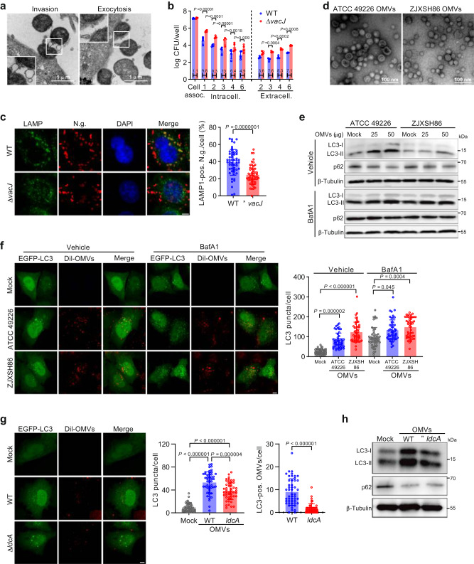
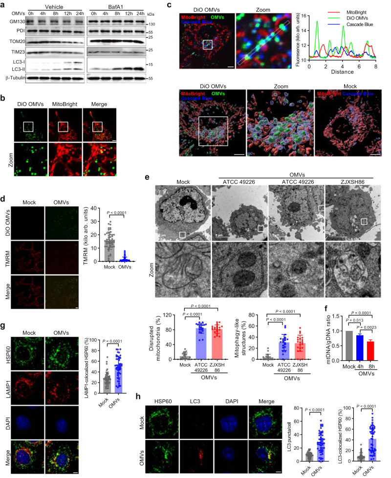
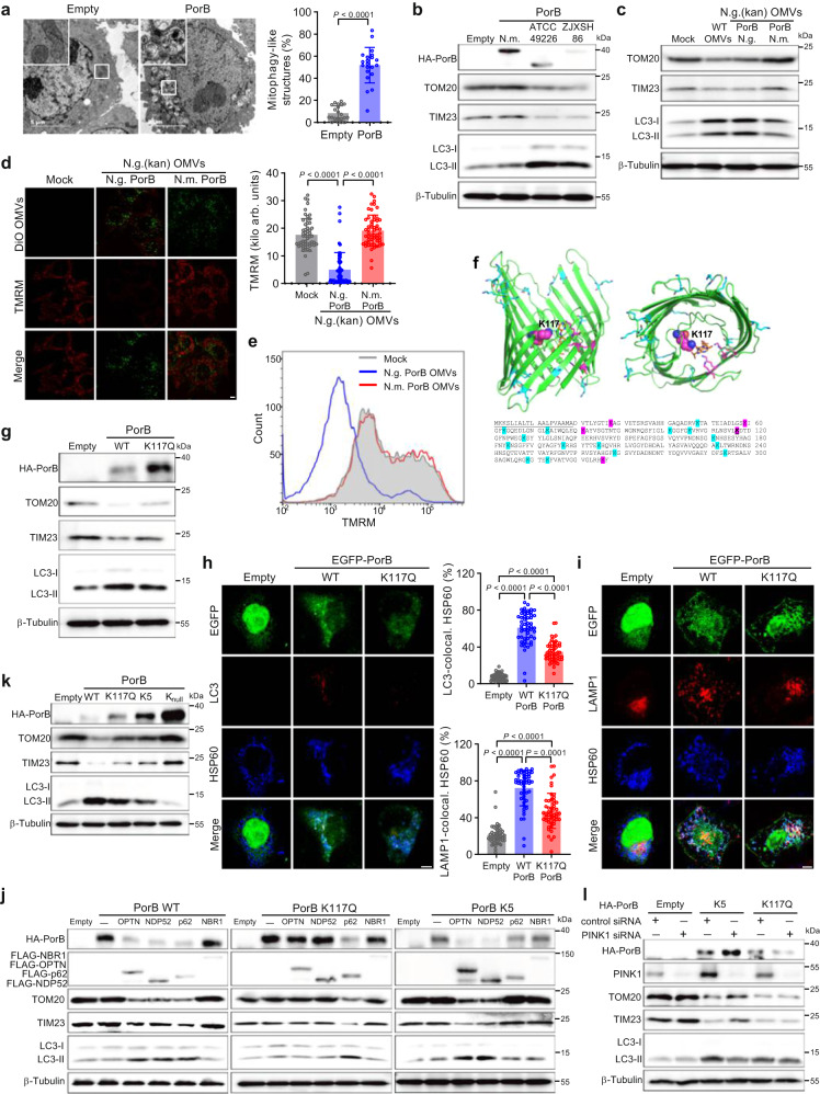
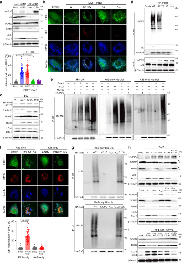
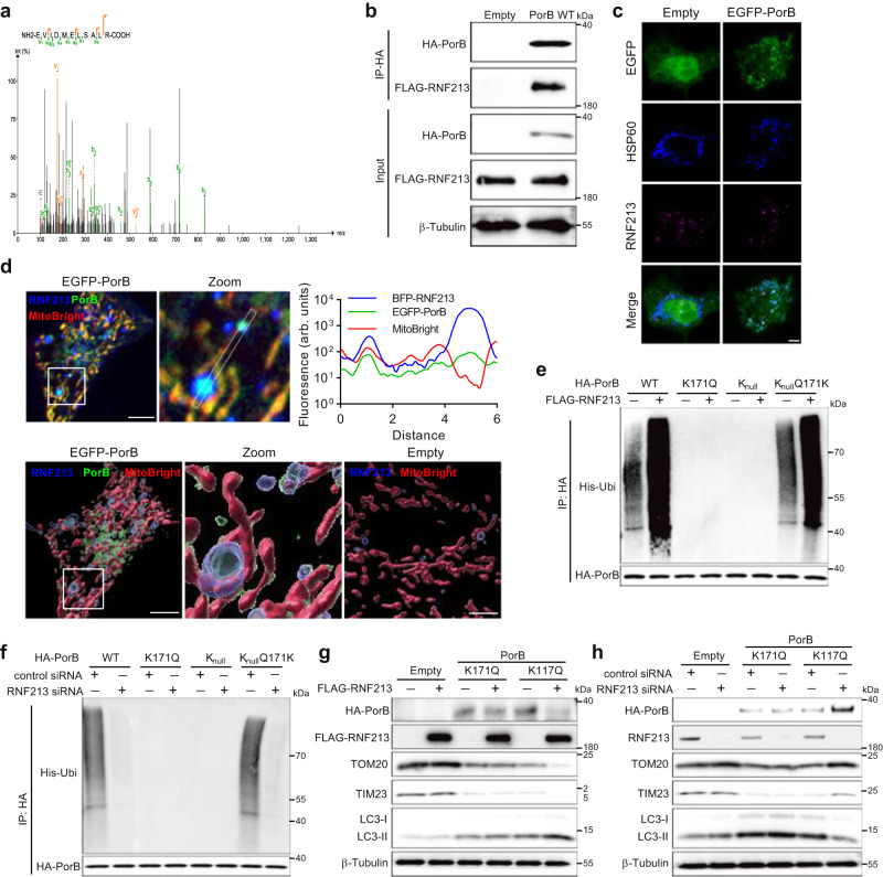
The bacterial pathogen Neisseria gonorrhoeae is able to invade epithelial cells and survive intracellularly. During this process, it secretes outer membrane vesicles (OMVs), however, the mechanistic details for interactions between gonococcal OMVs and epithelial cells and their impact on intracellular survival are currently not established. Here, we show that gonococcal OMVs induce epithelial cell mitophagy to reduce mitochondrial secretion of reactive oxygen species (ROS) and enhance intracellular survival. We demonstrate that OMVs deliver PorB to mitochondria to dissipate the mitochondrial membrane potential, resulting in mitophagy induction through a conventional PINK1 and OPTN/NDP52 mechanism. Furthermore, PorB directly recruits the E3 ubiquitin ligase RNF213, which decorates PorB lysine residue 171 with K63-linked polyubiquitin to induce mitophagy in a p62-dependent manner. These results demonstrate a mechanism in which polyubiquitination of a bacterial virulence factor that targets mitochondria directs mitophagy processes to this organelle to prevent its secretion of deleterious ROS.


Conflict of interest statement

The authors declare no competing interests.

Figures

Fig. 1
Fig. 1. Gonococcal OMVs induce autophagy flux in epithelial cells.
a TEM of gonococcal OMV secretion during invasion and exocytosis of HeLa cells. Images are representative of 2 independent experiments. b Enhanced intracellular survival and exocytosis of gonococcal ΔvacJ mutant in gentamicin protection assays with HeLa cells. Data are mean ± s.d. of log-normalized colony forming units (CFU) per well and relative differences in survival between WT and ΔvacJ are provided within the bars; n = 4 independent experiments, two-way ANOVA with posthoc Bonferroni test. c ΔvacJ mutant shows reduced LAMP1-colocalization after a one-hour challenge of HeLa cells. Scale bar, 5 μm. Data are mean ± s.d.; n = 53 cells, unpaired two-tailed t-test. d TEM images of purified gonococcal OMVs. Images are representative of 4 (ATCC 49226) and 3 (ZJXSH86) independent experiments. e LC3 Western blots show induction of autophagy flux in HeLa cells stimulated with gonococcal OMVs from strains ATCC 49226 and ZJXSH86. f Gonococcal OMVs induce accumulation of LC3 puncta in HeLa cells. Scale bar, 5 μm. Data are mean ± s.d.; n = 50 cells, Kruskal–Wallis with posthoc Dunn test (Vehicle Mock-ZJXSH86 OMVs: P < 10−15). g OMVs from the gonococcal ΔldcA mutant induce accumulation of LC3 puncta in HeLa cells, but largely lost colocalization with LC3 puncta. Scale bar, 5 μm. Data are mean ± s.d.; n = 50 cells, one-way ANOVA with post-hoc Tukey test for quantification of LC3 puncta (Mock-WT OMVs: P = 3 × 10−14; Mock-ΔldcA OMVs: P = 6 × 10−14), unpaired two-tailed t-test for quantification of LC3-positive OMVs (P = 5 × 10−13). h LC3 Western blots of OMV-stimulated HeLa cells show reduced accumulation of LC3 for the ΔldcA mutant. Cells in c, f, g are from 3 independent experiments. Western blots in e, h are representative of 3 independent experiments. Source data are provided as a Source Data file.
Fig. 2
Fig. 2. Gonococcal OMVs target mitochondria and induce mitophagy.
a Western blots of OMV-stimulated HeLa cells show specific autophagosome/lysosome-dependent degradation of mitochondrial marker proteins (TOM20 and TIM23), but not of Golgi (GM130) or endoplasmic reticulum (PDI). Western blots are representative of 3 independent experiments. b DiO-labeled gonococcal OMVs from strain ATCC 49226 colocalize with MitoBright-labeled mitochondria in HeLa cells. Scale bar, 5 μm. c Live HeLa cell microscopy and 3D image reconstruction shows OMVs remain associated with Cascade Blue-labeled endosomes when delivered to MitoBright-labeled mitochondria. Scale bar, 5 μm. The fluorescence colocalization profile of the line is shown. d Gonococcal OMVs from strain ATCC 49226 dissipate the mitochondrial membrane potential (MMP) in HeLa cells. Scale bar, 5 μm. Data are mean ± s.d.; n = 50 cells, unpaired two-tailed t-test, P < 10−15. e TEM of HeLa cells stimulated with gonococcal OMVs from strains ATCC 49226 and ZJXSH86 show mitochondrial disruption and capture in mitophagy-like structures. Data are mean ± s.d.; n = 21 cells, one-way ANOVA with post-hoc Tukey test for mitochondrial disruption (Mock-ATCC 49226 OMVs: P = 2 × 10−11; Mock-ZJXSH86 OMVs: P = 2 × 10−11), Kruskal–Wallis with posthoc Dunn test for mitochondria in mitophagy-like structures (Mock-ATCC 49226 OMVs: P = 1 × 10−6; Mock-ZJXSH86 OMVs: P = 7 × 10−6). f Quantitative real-time PCR showing a reduced mitochondrial to genomic DNA ratio in ATCC 49226 OMV-stimulated HeLa cells. Data are mean ± s.d.; n = 3 independent biological replicates, one-way ANOVA with post-hoc Tukey test. g Increased HSP60 and LAMP1 colocalization in ATCC 49226 OMV-stimulated HeLa cells. Scale bar, 5 μm. Data are mean ± s.d.; n = 50 cells, unpaired two-tailed t-test, P = 6 × 10−9. h Increased HSP60 and LC3 colocalization in ATCC 49226 OMV-stimulated HeLa cells. Scale bar, 5 μm. Data are mean ± s.d.; n = 50 cells, two-tailed Mann–Whitney test (LC3 puncta Mock-OMVs: P = 7 × 10−12; LC3 colocalized HSP60 Mock-ZJXSH86 OMVs: P = 4 × 10−15). Cells in d, e, g, h are from 3 independent experiments. Images in b, c are representative of 3 independent experiments. Source data are provided as a Source Data file.
Fig. 3
Fig. 3. Gonococcal OMVs deliver PorB to mitochondria to induce mitophagy.
a TEM of HeLa cells expressing gonococcal PorB from strain ATCC 49226 show mitochondrial capture in mitophagy-like structures. Data are mean ± s.d.; n = 21 cells, two-tailed Mann–Whitney test, P = 2 × 10−10. b Western blots showing degradation of mitochondrial proteins TOM20 and TIM23 in HeLa cells expressing gonococcal PorB from strains ATCC 49226 and ZJXSH86, but not for PorB from Neisseria mucosa. c Western blots showing that gonococcal OMVs expressing PorB from N. mucosa lost the ability to induce degradation of TOM20 and TIM23 in HeLa cells. d Gonococcal OMVs expressing PorB from N. mucosa lost the ability to dissipate the mitochondrial membrane potential (MMP) in HeLa cells. Scale bar, 5 μm. Data are mean ± s.d.; n = 55 cells from 4 independent experiments, Kruskal–Wallis with posthoc Dunn test (Mock-N.g. PorB: P = 8 × 10−13; N.g. PorB-N.m. PorB: P < 10−15). e Flow cytometry analysis of TMRM fluorescence intensity in HeLa cells stimulated with gonococcal OMVs expressing gonococcal PorB or PorB from N. mucosa. f Gonococcal PorB structure (pdb entry 4AUI) and sequence of PorB from strain ATCC 49226. Lysines are indicated in blue, or in magenta when located within the PorB channel and associated with ATP (orange) binding. g Western blots showing reduced degradation of TOM20 and TIM23 in HeLa cells expressing PorB K117Q. h Reduced HSP60 and LC3 colocalization in HeLa cells expressing PorB K117Q compared with PorB WT. Scale bar, 5 μm. Data are mean ± s.d.; n = 51 cells, Kruskal–Wallis with posthoc Dunn test (Empty-WT PorB: P < 10−15; Empty-PorB K117Q: P = 2 × 10−9; WT PorB-PorB K117Q: P = 1 × 10−5). i Reduced HSP60 and LAMP1 colocalization in HeLa cells expressing PorB K117Q compared with PorB WT. Scale bar, 5 μm. Data are mean ± s.d.; n = 51 cells, Kruskal–Wallis with posthoc Dunn test (Empty-WT PorB: P < 10−15; Empty-PorB K117Q: P = 9 × 10−8). j Western blots of HeLa 4KO cells showing p62 expression restores PorB K117Q-dependent degradation of TOM20 and TIM23, while expression of OPTN or NDP52 restores PorB K5-dependent degradation of TOM20 and TIM23. k Western blots showing impaired degradation of TOM20 and TIM23 in HeLa cells expressing PorB K117Q, K5 or Knull. l Western blots showing that knock-down of PINK1 in HeLa cells inhibits PorB K5-dependent degradation of TOM20 and TIM23, but not for PorB K117Q. Cells in a, h, i are from 3 independent experiments. Western blots in b, c, g, j, k, l are representative of 3 independent experiments. Source data are provided as a Source Data file.
Fig. 4
Fig. 4. Lysine 63-linked polyubiquitination of PorB induces p62-dependent mitophagy.
a Western blots showing that knock-down of p62 in HeLa cells inhibits PorB K117Q-dependent degradation of TOM20 and TIM23. b Reduced p62 and HSP60 colocalization in HeLa cells expressing PorB K5 or Knull compared with PorB WT. Scale bar, 5 μm. Data are mean ± s.d.; n = 50 cells, Kruskal–Wallis with posthoc Dunn test (Empty-WT PorB: P < 10−15; WT PorB-PorB K5: P < 10−15; WT PorB-PorB Knull: P < 10−15). c Western blots of HeLa 4KO cells expressing WT or truncated p62 show that the p62 LIR domain and UBI domain are indispensable for PorB K117Q-induced degradation of TOM20 and TIM23. d Western blots showing co-immunoprecipitation of ubiquitin and p62 after immunoprecipitation of PorB WT and PorB K117Q. e Western blots showing co-immunoprecipitation of both K63-linked and K48-linked polyubiquitin, after immunoprecipitation of PorB from HeLa cells, which is enhanced with inhibitors for autolysosomal degradation (BafA1, 3-MA) for K63-linked ubiquitin or proteasomal degradation (MG132) for K48-linked ubiquitin. f Colocalization between HSP60 and K63-linked polyubiquitin, but not K48-linked polyubiquitin, is induced in HeLa cells expressing PorB. Scale bar, 5 μm. Data are mean ± s.d.; n = 50 cells, two-tailed Mann–Whitney test, K63 only Empty-PorB K117Q: P < 10−15. g Western blots after immunoprecipitation of HA-PorB from HeLa cells show co-immunoprecipitation of K63-linked polyubiquitin is dependent on PorB lysine 171 and co-immunoprecipitation of K48-linked polyubiquitin is dependent on PorB lysine 128. h Western blots showing PorB-induced degradation of TOM20 and TIM23 in HeLa cells is dependent on PorB lysines 117 and 171. i Western blots showing gonococcal OMVs expressing PorB K117Q/K171Q lost the ability to induce degradation of TOM20 and TIM23. Cells in b and f are from 3 independent experiments. Western blots in a, c, d, e, g, h, i are representative of 3 independent experiments. Source data are provided as a Source Data file.
Fig. 5
Fig. 5. E3 ligase RNF213 interacts with PorB and catalyzes polyubiquitination.
a MS/MS spectrum of RNF213 peptide identified by mass spectrometry analysis of proteins co-immunoprecipitated with PorB from HeLa cells. b Western blots after immunoprecipitation of PorB from HeLa cells show co-immunoprecipitation of RNF213. c HeLa cells expressing PorB show colocalization between PorB, HSP60 and RNF213. Scale bar, 5 μm. d Live HeLa cell microscopy and 3D image reconstruction shows RNF213 surrounding PorB on MitoBright-labeled mitochondria. Scale bar, 5 μm. The fluorescence colocalization profile of the line is shown. e Western blots showing PorB K171-dependent enhanced co-immunoprecipitation of ubiquitin from HeLa cells transfected with an RNF213 expression vector. f Western blots showing that transfection of HeLa cells with RNF213 siRNA abolishes PorB K171-dependent co-immunoprecipitation of ubiquitin. g Western blots showing PorB K117Q, but not PorB K171Q, enhances PorB-induced degradation of TOM20 and TIM23 in HeLa cells transfected with an RNF213 expression vector. h Western blots showing that transfection of HeLa cells with RNF213 siRNA prevents PorB-induced degradation of TOM20 and TIM23 for PorB K117Q, while degradation remains unaffected for PorB K171Q. Western blots in b, eh are representative of 3 independent experiments. Images in c and d are representative of 3 independent experiments. Source data are provided as a Source Data file.
Fig. 6
Fig. 6. OMV-induced mitophagy reduces generation of mitochondrial ROS to enhance intracellular survival.
a Live-cell microscopy showing that OMVs transiently induce generation of mitochondrial ROS in HeLa cells. Scale bar, 5 μm. Data are mean ± s.d.; n = 50 cells, two-way ANOVA with posthoc Bonferroni test, P < 10−15 for all reported values. b Live-cell microscopy showing that inhibition of mitophagy with Mdivi-1 prolongs mitochondrial generation of ROS in OMV-stimulated HeLa cells. Data are mean ± s.d.; n = 50 cells, two-way ANOVA with posthoc Bonferroni test, Vehicle-Mdivi-1: P < 10−15 for 3 and 6 h. c Inhibition of mitophagy with Mdivi-1 reduces gonococcal intracellular survival in gentamicin protection assays, while activation of mitophagy with CCCP or scavenging of mitochondrial ROS with mito-TEMPO enhances intracellular survival. Data are mean ± s.d.; n = 4, two-way ANOVA with posthoc Bonferroni test, Vehicle-mito-TEMPO at 3 h: P = 4 × 10−11, Vehicle-CCCP at 6 h: P = 1 × 10−14, Vehicle-mito-TEMPO at 6 h: P < 10−15. d Prior stimulation of HeLa cells with gonococcal WT OMVs to induce mitophagy enhances gonococcal intracellular survival in gentamicin protection assays, while OMVs from gonococcal mutants expressing PorB from N. mucosa or PorB K117Q/K171Q are unable to enhance intracellular survival. Data are mean ± s.d.; n = 4, two-way ANOVA with posthoc Bonferroni test. e Prior stimulation of Mdivi-1-pretreated HeLa cells with gonococcal OMVs reduces gonococcal intracellular survival in gentamicin protection assays. Data are mean ± s.d.; n = 4, two-way ANOVA with posthoc Bonferroni test, OMVs-OMVs+Mdivi-1 at 2 h: P = 9 × 10−11, Vehicle-OMVs+Mdivi1 at 4 h: P = 2 × 10−7, OMVs-OMVs+Mdivi-1 at 4 h: P < 10−15. f Live-cell microscopy of gonococcal-challenged HeLa cells showing reduced generation of mitochondrial ROS for the ΔvacJ mutant and enhanced generation of mitochondrial ROS for N. gonorrhoeae expressing PorB from N. mucosa or PorB K117Q/K171Q. Data are mean ± s.d.; n = 50 cells, two-way ANOVA with posthoc Bonferroni test, WT- ΔvacJ at cell associated: P < 10−15, WT-ΔvacJ at 3 h: P = 1 × 10−13, WT-N.m. PorB at 3 h: P < 10−15, WT-PorB K117Q/K171Q at 3 h: P < 10−15, WT-N.m. PorB at 6 h: P = 4 × 10−12, WT-PorB K117Q/K171Q at 6 h: P = 6 × 10−14. g Gonococcal mutants expressing PorB from N. mucosa or PorB K117Q/K171Q show reduced intracellular survival in gentamicin protection assays. Data are mean ± s.d.; n = 4, two-way ANOVA with posthoc Bonferroni test. Cells in a, b, f are from 3 independent experiments. Data in ce, g are log-normalized CFU per well from 4 independent experiments, with fold-changes in survival compared with the Vehicle or Mock provided within the bars. Source data are provided as a Source Data file.
Fig. 7
Fig. 7. Summary illustration of the gonococcal OMV-induced mitophagy mechanism through a dual PorB-dependent pathway.
Gonococcal OMVs are endocytosed by epithelial cells and deliver PorB to the mitochondria. Insertion of PorB in the mitochondrial membrane dissipates the mitochondrial membrane potential (MMP). Dissipation of MMP is dependent on ATP binding by lysines in the PorB channel, such as lysine 117, that prolong the channel in an open position. Dissipation of MMP results in PINK1- and OPTN/NDP52-dependent activation of mitophagy. PorB is furthermore decorated by K63-linked polyubiquitin chains at PorB lysine 171 through activity of E3 ubiquitin ligase RNF213, which activates mitophagy in a p62-dependent manner. OMV- and PorB-dependent activation of mitophagy abolishes mitochondrial generation of reactive oxygen species (ROS) upon gonococcal invasion of epithelial cells, which results in enhanced intracellular survival. Figure created with cartoon components by Figdraw [www.figdraw.com].

References

    1. Muenzner P, Bachmann V, Zimmermann W, Hentschel J, Hauck CR. Human-restricted bacterial pathogens block shedding of epithelial cells by stimulating integrin activation. Science. 2010;329:1197–1201. doi: 10.1126/science.1190892. - DOI - PubMed
    1. Muenzner P, Hauck CR. Neisseria gonorrhoeae blocks epithelial exfoliation by nitric-oxide-mediated metabolic cross talk to promote colonization in mice. Cell Host Microbe. 2020;27:793–808. doi: 10.1016/j.chom.2020.03.010. - DOI - PubMed
    1. Yu Q, et al. Neisseria gonorrhoeae infects the heterogeneous epithelia of the human cervix using distinct mechanisms. PLoS Pathog. 2019;15:e1008136. doi: 10.1371/journal.ppat.1008136. - DOI - PMC - PubMed
    1. Faulstich M, Bottcher JP, Meyer TF, Fraunholz M, Rudel T. Pilus phase variation switches gonococcal adherence to invasion by caveolin-1-dependent host cell signaling. PLoS Pathog. 2013;9:e1003373. doi: 10.1371/journal.ppat.1003373. - DOI - PMC - PubMed
    1. Rechner C, Kuhlewein C, Muller A, Schild H, Rudel T. Host glycoprotein Gp96 and scavenger receptor SREC interact with PorB of disseminating Neisseria gonorrhoeae in an epithelial invasion pathway. Cell Host Microbe. 2007;2:393–403. doi: 10.1016/j.chom.2007.11.002. - DOI - PubMed

MeSH terms

LinkOut - more resources