. 2024 Feb 23;4(1):29.
doi: 10.1038/s43856-024-00452-8.

Multi-scale V-net architecture with deep feature CRF layers for brain extraction

Affiliations

Multi-scale V-net architecture with deep feature CRF layers for brain extraction

Jong Sung Park et al. Commun Med (Lond). .

Abstract

Background: Brain extraction is a computational necessity for researchers using brain imaging data. However, the complex structure of the interfaces between the brain, meninges and human skull have not allowed a highly robust solution to emerge. While previous methods have used machine learning with structural and geometric priors in mind, with the development of Deep Learning (DL), there has been an increase in Neural Network based methods. Most proposed DL models focus on improving the training data despite the clear gap between groups in the amount and quality of accessible training data between.

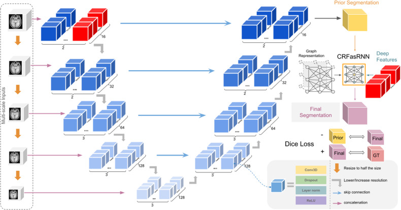
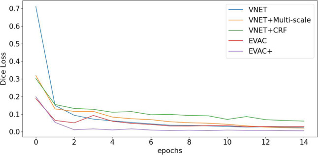
Methods: We propose an architecture we call Efficient V-net with Additional Conditional Random Field Layers (EVAC+). EVAC+ has 3 major characteristics: (1) a smart augmentation strategy that improves training efficiency, (2) a unique way of using a Conditional Random Fields Recurrent Layer that improves accuracy and (3) an additional loss function that fine-tunes the segmentation output. We compare our model to state-of-the-art non-DL and DL methods.

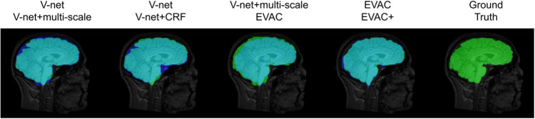
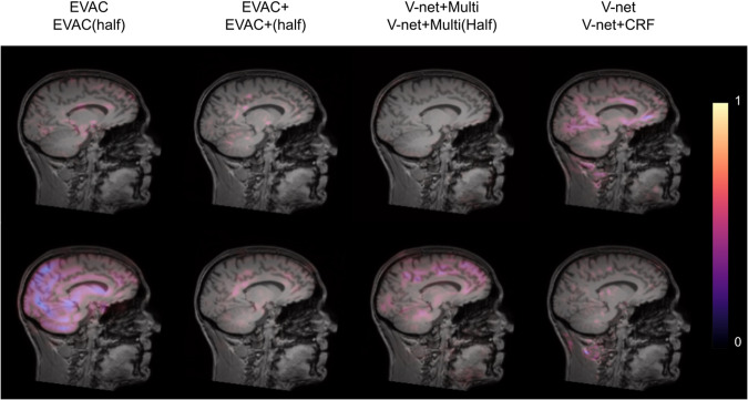
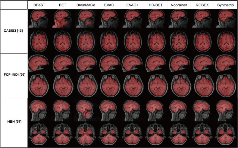
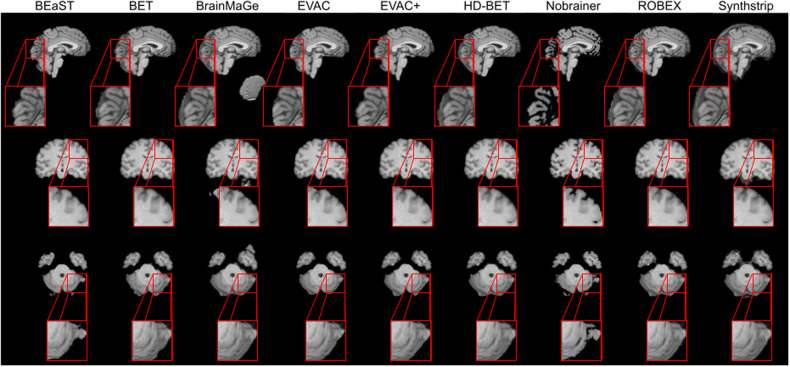
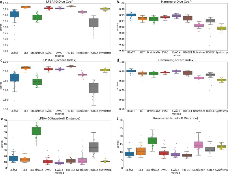
Results: Results show that even with limited training resources, EVAC+ outperforms in most cases, achieving a high and stable Dice Coefficient and Jaccard Index along with a desirable lower Surface (Hausdorff) Distance. More importantly, our approach accurately segmented clinical and pediatric data, despite the fact that the training dataset only contains healthy adults.

Conclusions: Ultimately, our model provides a reliable way of accurately reducing segmentation errors in complex multi-tissue interfacing areas of the brain. We expect our method, which is publicly available and open-source, to be beneficial to a wide range of researchers.

Plain language summary

Computational processing of brain images can enable better understanding and diagnosis of diseases that affect the brain. Brain Extraction is a computational method that can be used to remove areas of the head that are not the brain from images of the head. We compared various different computational methods that are available and used them to develop a better method. The method we describe in the paper is more accurate at imaging the brain of both healthy individuals and those known to have diseases that affect the brain than the other methods we evaluated. Our method might enable better understanding and diagnosis of diseases that affect the brain in the future.


Conflict of interest statement

The authors declare no competing interests.

Figures

Fig. 1
Fig. 1. Summary of the model architecture.
The architecture uses V-net as its base model with the following important changes: multi-resolutional raw inputs, modified CRFasRNN and additional Dice Loss. The CRF layer uses the rear layer of the first level of the encoder. A negative Dice Loss is calculated between the base model’s output and CRF layer’s output.
Fig. 2
Fig. 2. Qualitative ablation study.
Blue represents a model without a certain change, green the model with one of the proposed changes and teal the overlapping regions. Ground truth is added for reference. We can see that while both multi-scale inputs and conditional random fields do a great job of recovering False Negatives and removing False Positives, a combination of them gives a better segmentation. The additional loss function also fine-tunes the output closer to the ground truth.
Fig. 3
Fig. 3. Dice Loss comparison for ablation study.
The original Dice Loss plot. The values in the plot do not include the proposed regularizing negative Dice Loss of our model. It clearly shows an efficiency increase in training for each improvement. Note that our model (EVAC+) with the additional Dice Loss trains better even in terms of the original Dice Loss.
Fig. 4
Fig. 4. Important image features highlighted through integrated gradients.
Larger value indicates a larger gradient within that region, suggesting higher importance in prediction. Note that our model is not affected by most non-brain regions such as dura mater and neck regions. The figure also shows that EVAC+ has less important features within the brain, suggesting that the proposed CRF layer is contributing more to the model.
Fig. 5
Fig. 5. Qualitative robustness analysis of the compared methods.
The images are from OASIS, Parkinson’s dataset from the FCP-INDI project and pediatric brains from the Healthy Brain Network dataset. Red marks the segmentation results of each method.
Fig. 6
Fig. 6. Qualitative comparison between models.
The image shows comparison between our models with (EVAC+) and without the additional Dice Loss (EVAC) and other established state-of-the-art models. Specific regions were zoomed in to emphasize the improvements our model is achieving. Results show that our models get an accurate local segmentation on the surface, whereas most of the other methods either under-segment or include the dura mater. Similar accuracy improvements are also visible near the central sulcus and the cerebellum. T1 images from the IXI dataset.
Fig. 7
Fig. 7. Quantitative analysis between models.
The plots indicate (a) Dice Coefficient, (c) Jaccard Index, and (e) Hausdorff Distance for the LPBA40 dataset (n = 39) and (b) Dice Coefficient, (d) Jaccard Index and (f) Hausdorff Distance for the Hammers Atlas dataset (n = 30). Our models with (EVAC+) and without the additional Dice Loss (EVAC) have a stable near-top accuracy in both datasets and metrics, while others have either lower scores in a dataset or unstable results.

References

    1. Rehman HZU, Hwang H, Lee S. Conventional and deep learning methods for skull stripping in brain MRI. Appl. Sci. 2020;10:1773. doi: 10.3390/app10051773. - DOI
    1. Smith SM. Fast robust automated brain extraction. Human Brain Mapping. 2002;17:143–155. doi: 10.1002/hbm.10062. - DOI - PMC - PubMed
    1. Atkins, M. S., Siu, K., Law, B., Orchard, J. J. & Rosenbaum, W. L. Difficulties of t1 brain MRI segmentation techniques. In: Medical Imaging 2002: Image Processing (eds Milan, S., J & Michael, F.), 1837–1844 (SPIE, 2002).
    1. Cox RW. Afni: what a long strange trip it’s been. Neuroimage. 2012;62:743–747. doi: 10.1016/j.neuroimage.2011.08.056. - DOI - PMC - PubMed
    1. Ségonne F, et al. A hybrid approach to the skull stripping problem in MRI. Neuroimage. 2004;22:1060–1075. doi: 10.1016/j.neuroimage.2004.03.032. - DOI - PubMed