

A Rare Cause of Deep Vein Thrombosis in a Young Orchestra Conductor
- PMID: 38396393
- PMCID: PMC10887723
- DOI: 10.3390/diagnostics14040354
A Rare Cause of Deep Vein Thrombosis in a Young Orchestra Conductor
Abstract
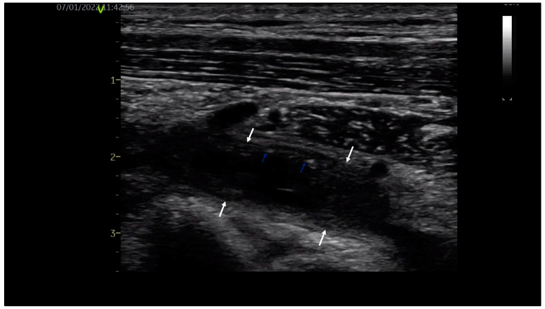
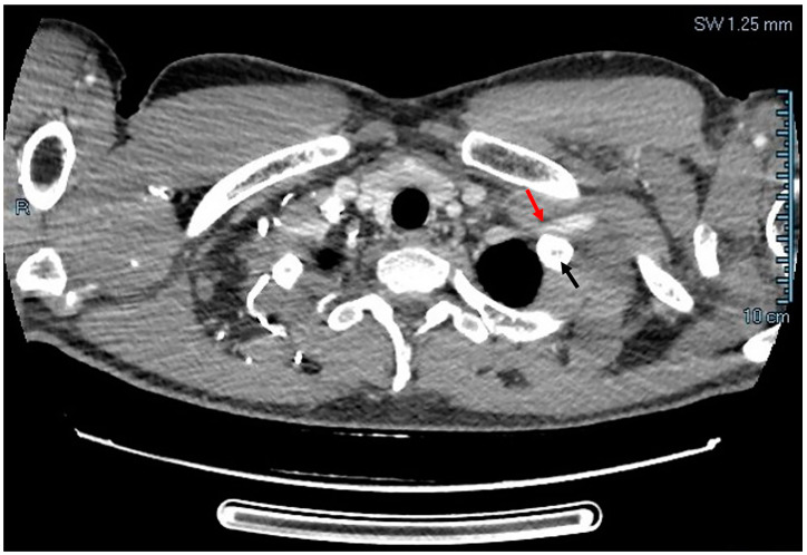
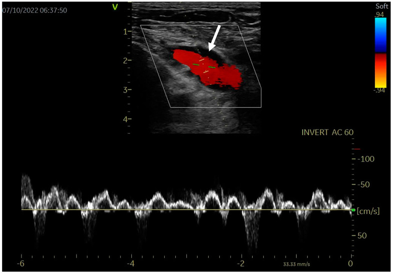
Upper extremity deep vein thrombosis (DVT) of the axillary/subclavian veins is rare (5-10% of DVT). After clinical suspicion and duplex ultrasound, anticoagulation, surgical decompression and sometimes thrombolysis are mandatory due to complications. We discuss the case of a young healthy orchestra conductor with primary DVT of the left upper extremity and concomitant left shoulder musculo-tendinous traumatic injury. Symptoms of both conditions and subtle signs of upper extremity DVT delayed the diagnosis until full-blown DVT occurred. After successful anticoagulation and surgical TOS (thoracic outlet syndrome) decompression, evolution was favorable, without recurrent thrombosis.
Keywords: anticoagulants; effort-induced upper extremity deep vein thrombosis; surgical decompression; thoracic outlet syndrome.
Conflict of interest statement
The authors declare no conflicts of interest.
Figures
References
-
- Kastora S.L., Oduyoye O., Mahmood S. Upper extremity deep venous thrombosis prevalence in the NHS Grampian Medical Ambulatory clinic: Diagnostic, therapeutic, and prognostic considerations in oncology patients. Irish J. Med. Sci. 2022;191:1569–1575. doi: 10.1007/s11845-021-02775-0. - DOI - PMC - PubMed
LinkOut - more resources
Full Text Sources
