Case Reports
. 2024 Feb 8;14(4):577.
doi: 10.3390/ani14040577.

Treatment of Medial Instability of the Carpometacarpal and Tarsometatarsal Joints Using the Isolock® System in Two Dogs

Affiliations
Case Reports

Treatment of Medial Instability of the Carpometacarpal and Tarsometatarsal Joints Using the Isolock® System in Two Dogs

Stefania Pinna et al. Animals (Basel). .

Abstract

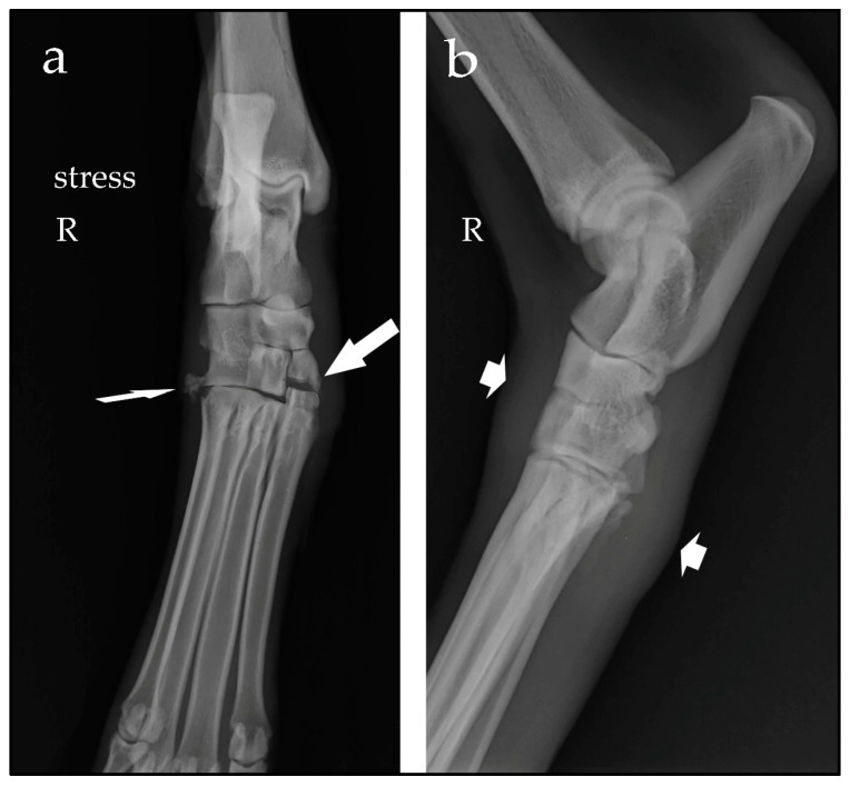
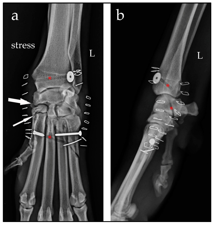
This case report describes a novel procedure using the Isolock Intrauma® implant system for treating medial instability of the carpometacarpal and tarsometatarsal joints, as demonstrated in in two dogs. A 9-year-old spayed female Spanish greyhound presented with a non-weight-bearing right hindlimb following a trauma. The clinical and radiological findings confirmed medial tarsometatarsal instability consistent with valgus deviation of the tarsus and the opening of the joint line on the medial aspect from the first to the third tarsometatarsal joints. A 4-year-old female Drahthaar presented with a non-weight-bearing left forelimb, swelling of the carpus and valgus instability. Radiological examination revealed a widening of the spaces between the intermedioradial carpal bone, second carpal bone and metacarpal bone II, confirming the medial carpometacarpal instability. In both cases, the Isolock system, an implant including ultra-high-molecular-weight polyethylene suture (UHMWPE), was used to reinforce the medial joint structures. Minor short-term complications were observed, such as swelling of the tarsal surgical site and hyperextension of the carpus, but these resolved spontaneously. No lameness or major complications were reported five months postoperatively. Carpometacarpal and tarsometatarsal instabilities are rare diseases in dogs as compared to subluxations of the other joints of the carpus and tarsus. There are no previous reports regarding the use of a UHMPWE implant for the treatment of these rare joint injuries, though the present case report suggests the validity and efficacy of the Isolock Intrauma® implant for restoring carpal and tarsal stability and preserving joint mobility.

Keywords: carpometacarpal joint; dog; tarsometatarsal joint; valgus instability.


Conflict of interest statement

The authors declare no conflicts of interest.

Figures

Figure 1
Figure 1
Stressed dorsoplantar radiographic view (a) and mediolateral view (b) of the right (R) tarsus. Note the opening of the tarsometatarsal joint line between the first to the third distal tarsal bones and the base of the metatarsal bones I to III (large arrow), the avulsion fracture of the proximal base of metatarsal bone V (narrow arrow) and the soft-tissue swelling (short arrows).
Figure 2
Figure 2
Stressed dorsopalmar radiographic view (a) and mediolateral view (b) of the left (L) carpus taken at the time of diagnosis. Note the opening of the spaces between the intermedioradial carpal bone, second carpal bone and metacarpal bone II (narrow arrows). An oblique fracture of the body of metacarpal bone V is evident (large arrow).
Figure 3
Figure 3
Schematic drawing of the surgical procedure for the treatment of tarsometatarsal instability. The black arrows indicate the direction of the insertion of the Isolock implant through the tarsal and metatarsal tunnels and the introduction of the interference screw.
Figure 4
Figure 4
Schematic drawing of the surgical procedure for the treatment of carpometacarpal instability. The black arrows indicate the direction of the insertion of the Isolock implant through the carpal and metacarpal tunnels and the introduction of the interference screw.
Figure 5
Figure 5
Stressed dorsoplantar radiographic view (a) and mediolateral view (b) of the right (R) tarsus taken postoperatively. The tarsometatarsal joint space has been restored to normal (arrow). The tarsal and metatarsal tunnels are visible (red asterisks); note the titanium button and interference screw placed at the end of the UHMWPE tensioned suture (not visible radiologically).
Figure 6
Figure 6
Stressed dorsopalmar radiographic view (a) and mediolateral view (b) of the left (L) carpus taken immediately after surgery. The joint space between the intermedioradial carpal bone and the second carpal bone is still slightly increased (large arrow); the joint line between the second carpal bone and metacarpal bone II is normal (narrow arrow). The carpal and metacarpal tunnels are visible (red asterisks); the titanium button and interference screw are in the correct position. The lag screw and the K-wire are correctly positioned to reduce the fracture of metacarpal bone V.
Figure 7
Figure 7
Stressed dorsoplantar radiographic view (a) and mediolateral view (b) of the right (R) tarsus taken five months after surgery. Radiographic images show a partial pull-out of the screw, slight soft-tissue swelling, and mild amorphous periosteal reaction of the dorsomedial aspect of the tarsometatarsal joint (short arrows). A bony callus is visible and is consistent with the healing of the avulsion fracture at the base of metatarsal bone V (narrow arrow).
Figure 8
Figure 8
Stressed dorsopalmar radiographic view (a) and mediolateral view (b) of the left (L) carpus taken five months after surgery. Radiographic images show a nearly normal joint space, no sign of line fracture and a bony callus (narrow arrows). Mild soft-tissue swelling is noted (short arrows). No migration of the orthopaedic implants is observed.

Similar articles

References

    1. Carmichael S., Marshall W. Tarsus and metatarsus. In: Johnson S.A., Tobias K.M., editors. Veterinary Surgery Small Animal. 2nd ed. Volume 1. Elsevier Saunders; St. Louis, MO, USA: 2017. pp. 1193–1209.
    1. Kapatkin A.S., Garcia-Nolen T., Hayashi K. Carpus, metacarpus and digits. In: Johnson S.A., Tobias K.M., editors. Veterinary Surgery Small Animal. 2nd ed. Volume 1. Elsevier Saunders; St. Louis, MO, USA: 2017. pp. 920–938.
    1. Evans H.E., de Lahunta A., editors. Miller’s Anatomy of the Dog. 4th ed. Elsevier Saunders; St. Louis, MO, USA: 2013. Arthrology; pp. 158–181.
    1. Earley T.D., Dee J.F. Trauma to the carpus, tarsus, and phalanges of dogs and cats. Vet. Clin. N. Am. Small Anim. Pract. 1980;10:717–747. doi: 10.1016/S0195-5616(80)50063-X. - DOI - PubMed
    1. Vaughan L.C. Disorders of the tarsus in the dog. I. Br. Vet. J. 1987;143:388–401. doi: 10.1016/0007-1935(87)90016-9. - DOI - PubMed

Publication types

LinkOut - more resources